Anatomy ofthe Eye
1. The Eyeball
2. Ocular Adnexa
3.
THE EYEBALL
Theeyeball is a cystic structure situated in the orbit.
Eyeball is not a sphere but an OBLATE SPHEROID
OBLATE SPHEROID: it is slightly flattened at the poles and wider at the
equator.
Connected to the brain by the optic nerve.
protected by bony orbit and eyelids.
4.
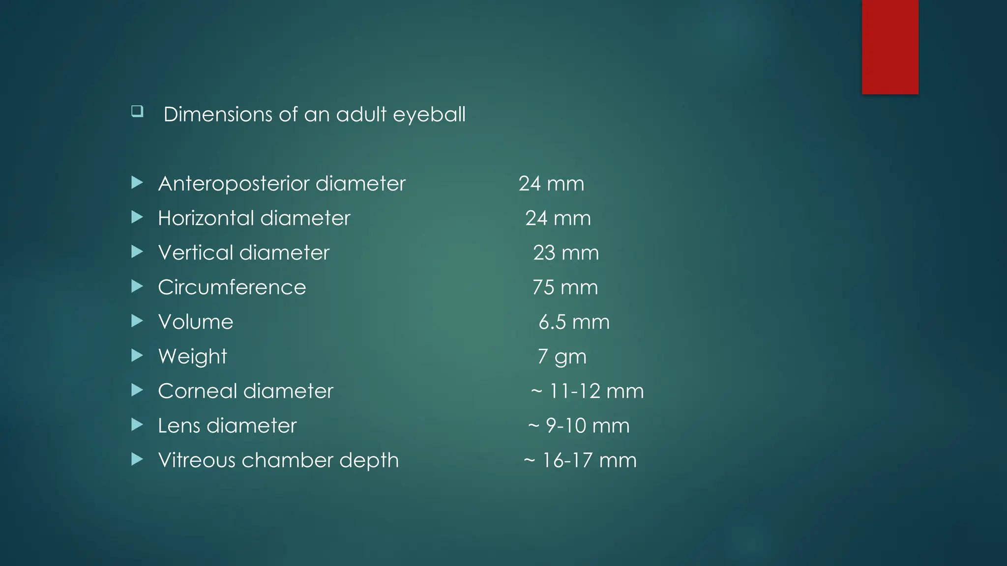
Dimensions ofan adult eyeball
Anteroposterior diameter 24 mm
Horizontal diameter 24 mm
Vertical diameter 23 mm
Circumference 75 mm
Volume 6.5 mm
Weight 7 gm
Corneal diameter ~ 11-12 mm
Lens diameter ~ 9-10 mm
Vitreous chamber depth ~ 16-17 mm
5.
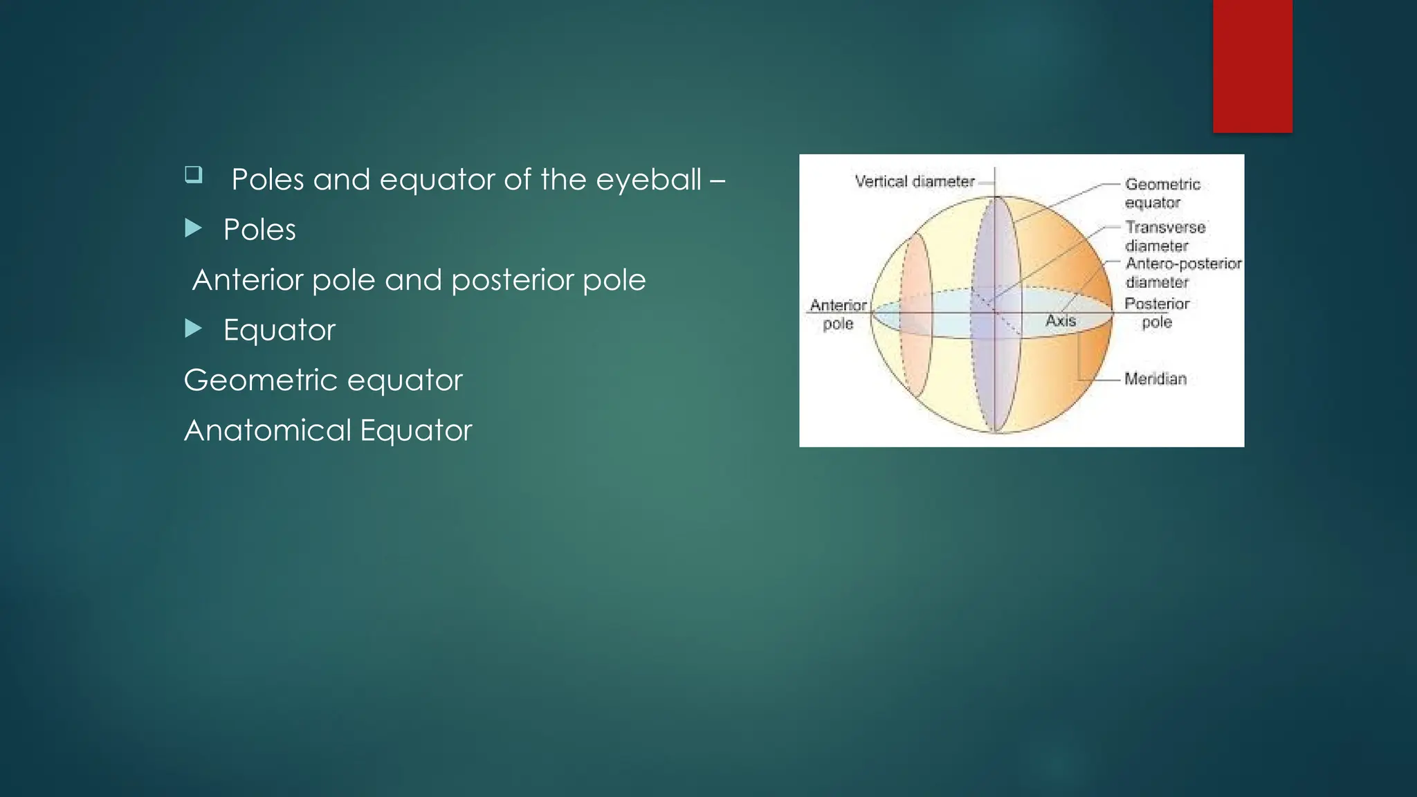
Poles andequator of the eyeball –
Poles
Anterior pole and posterior pole
Equator
Geometric equator
Anatomical Equator
1. Fibrous Coat
Its dense strong wall which protects the intraocular contents.
Anterior 1/6th
of the fibrous coat is transparent & its called cornea.
Posterior 5/6th
opaque part is called Sclera.
9.
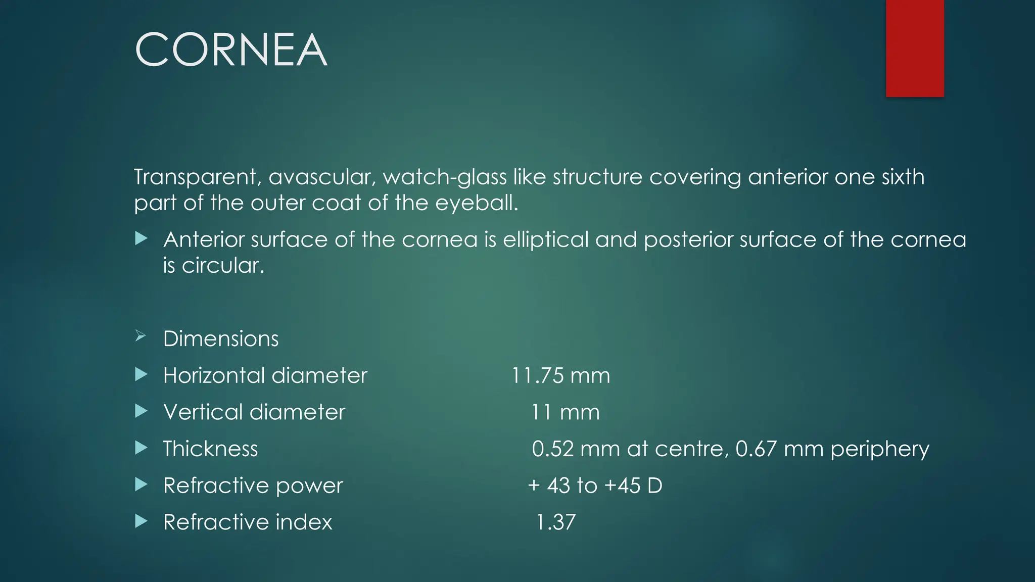
CORNEA
Transparent, avascular, watch-glasslike structure covering anterior one sixth
part of the outer coat of the eyeball.
Anterior surface of the cornea is elliptical and posterior surface of the cornea
is circular.
Dimensions
Horizontal diameter 11.75 mm
Vertical diameter 11 mm
Thickness 0.52 mm at centre, 0.67 mm periphery
Refractive power + 43 to +45 D
Refractive index 1.37
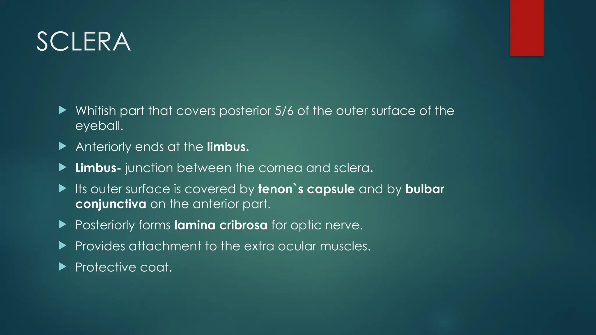
SCLERA
Whitish partthat covers posterior 5/6 of the outer surface of the
eyeball.
Anteriorly ends at the limbus.
Limbus- junction between the cornea and sclera.
Its outer surface is covered by tenon`s capsule and by bulbar
conjunctiva on the anterior part.
Posteriorly forms lamina cribrosa for optic nerve.
Provides attachment to the extra ocular muscles.
Protective coat.
12.
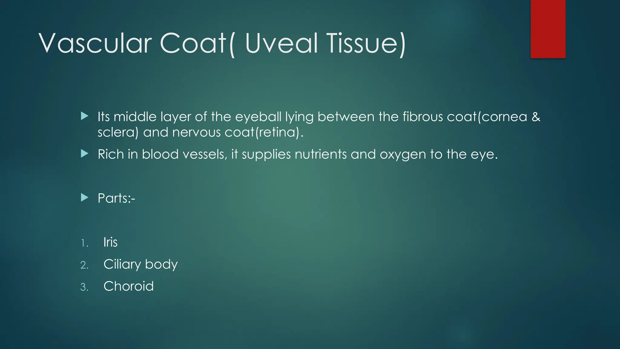
Vascular Coat( UvealTissue)
Its middle layer of the eyeball lying between the fibrous coat(cornea &
sclera) and nervous coat(retina).
Rich in blood vessels, it supplies nutrients and oxygen to the eye.
Parts:-
1. Iris
2. Ciliary body
3. Choroid
13.
IRIS
The irisis the coloured part of the eyeball.
Location-Lies between the cornea and the crystalline lens, in front of
the ciliary body.
Shape – thin circular disc with a central opening is called pupil
( 4mm).
Functions- controls the amount of the light entering into the eye by
adjusting the pupil size.
At periphery attaches to anterior surface of ciliary body.
Ciliary body
Middlelayer triangular shape of the uvea.
Location- between the iris ( anteriorly) and choroid (posteriorly).
Forward continuation of the choroid.
Aqueous humour formation and drainage.
Helps in in accommodation
Supports in lens via suspensory ligaments.
16.
Ciliary body consist5 layers-
1. Supraciliary lamina
2. Stroma of the ciliary body
3. Layer of pigmented epithelium
4. Layer of non-pigmented layer
5. Internal limiting layer
17.
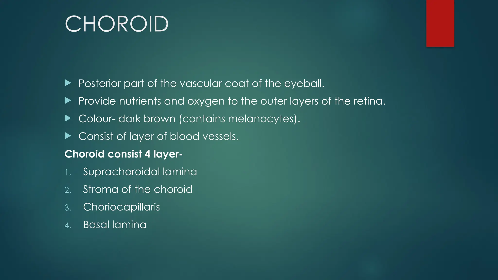
CHOROID
Posterior partof the vascular coat of the eyeball.
Provide nutrients and oxygen to the outer layers of the retina.
Colour- dark brown (contains melanocytes).
Consist of layer of blood vessels.
Choroid consist 4 layer-
1. Suprachoroidal lamina
2. Stroma of the choroid
3. Choriocapillaris
4. Basal lamina
18.
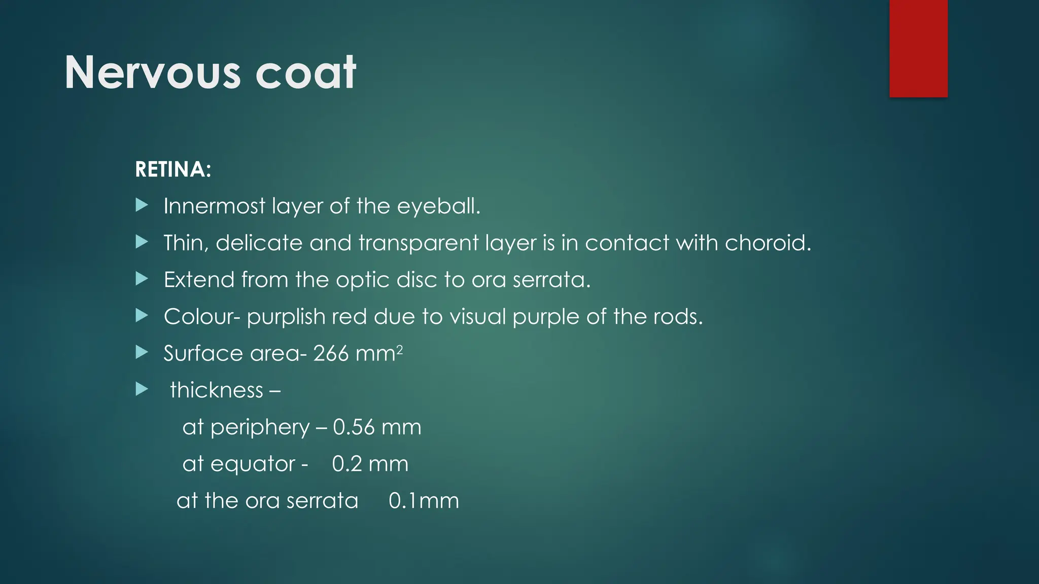
Nervous coat
RETINA:
Innermostlayer of the eyeball.
Thin, delicate and transparent layer is in contact with choroid.
Extend from the optic disc to ora serrata.
Colour- purplish red due to visual purple of the rods.
Surface area- 266 mm2
thickness –
at periphery – 0.56 mm
at equator - 0.2 mm
at the ora serrata 0.1mm
Segments of theeyeball
The eyeball is divided into 2 segments.
1. ANTERIOR SEGMENT
Location- in front of the lens
Structure anterior to the lens and ciliary body.
Anterior segment has two component-
A. Anterior chamber-
Between the cornea and iris, filled with aqueous humour.
About 2.5 mm in depth, and contains 2.5 ml of AH
B. Between the iris and lens, also filled with AH.
21.
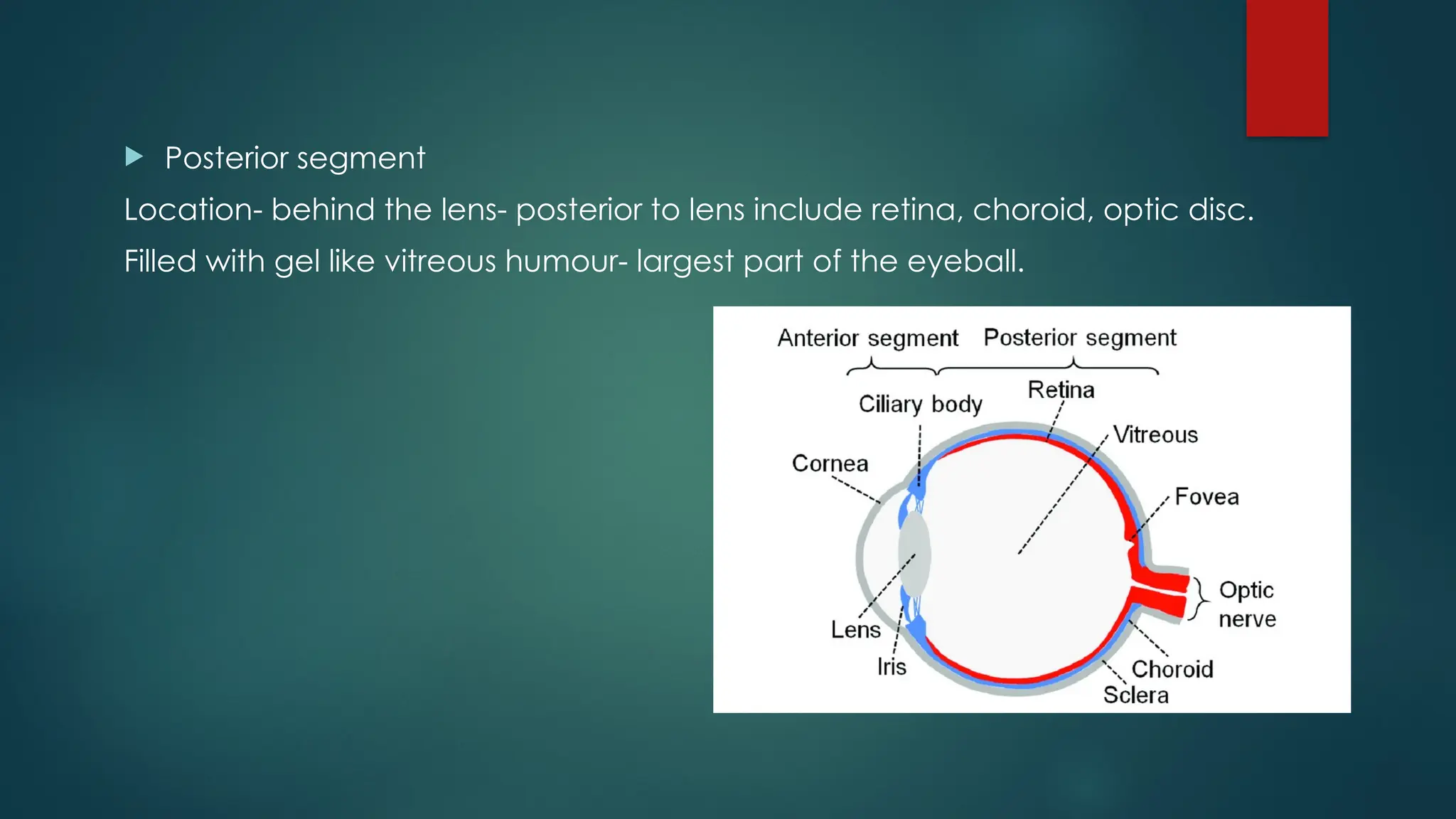
Posterior segment
Location-behind the lens- posterior to lens include retina, choroid, optic disc.
Filled with gel like vitreous humour- largest part of the eyeball.
22.
Crystalline lens
Biconvex
Suspended behind iris by zonules
Transparent, avascular, no nerves
Grows through the life
Structure- capsule, epithelium, cortex, nucleus.
Thickness- ~ 4 mm
Diameter- ~9-10 mm
Anterior radius of curvature- 10 mm, posterior curvature- 6 mm
Refractive power- 15-17 D
Refractive index- 1.39
23.
Ocular adnexa
Theadnexa are the accessory structures surrounding the eyeball, which
support and protect it.
They do not form part of the eyeball itself but are essential for proper eye
function.
Included structures that helps in protection, movement, lubricants and overall
ocular health.
A. The bony orbit
B. Eyelids
C. Conjunctiva
D. Lacrimal apparatus
E. Extraocular muscles
24.
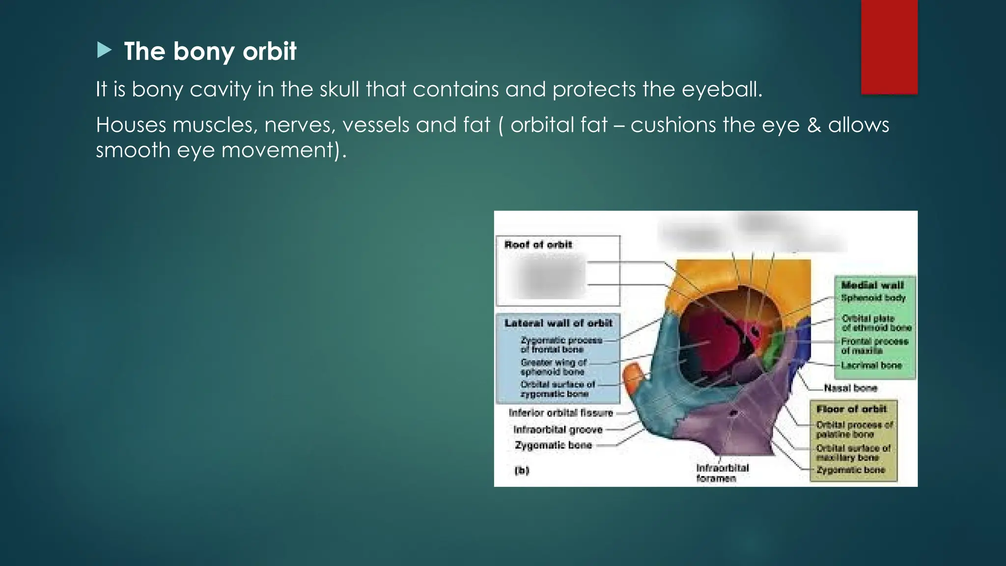
The bonyorbit
It is bony cavity in the skull that contains and protects the eyeball.
Houses muscles, nerves, vessels and fat ( orbital fat – cushions the eye & allows
smooth eye movement).
25.
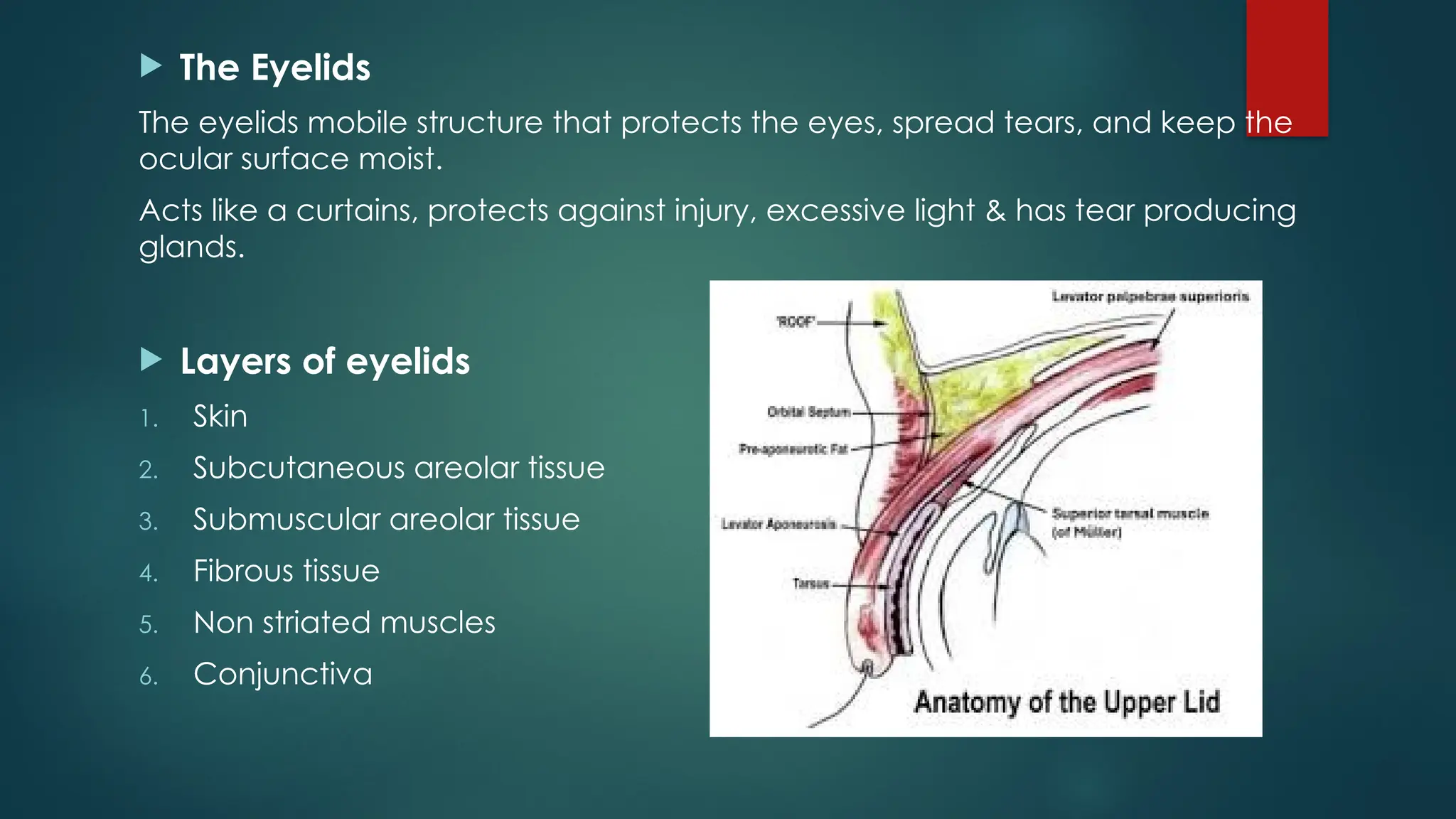
The Eyelids
Theeyelids mobile structure that protects the eyes, spread tears, and keep the
ocular surface moist.
Acts like a curtains, protects against injury, excessive light & has tear producing
glands.
Layers of eyelids
1. Skin
2. Subcutaneous areolar tissue
3. Submuscular areolar tissue
4. Fibrous tissue
5. Non striated muscles
6. Conjunctiva
26.
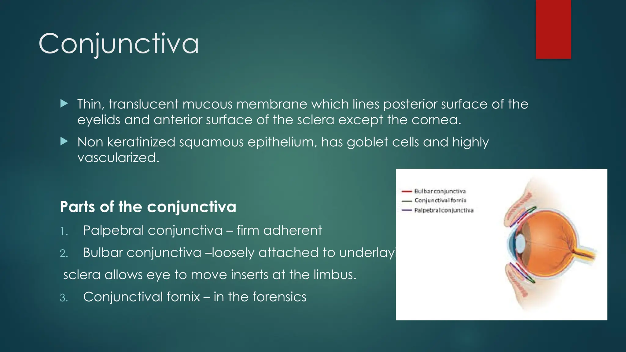
Conjunctiva
Thin, translucentmucous membrane which lines posterior surface of the
eyelids and anterior surface of the sclera except the cornea.
Non keratinized squamous epithelium, has goblet cells and highly
vascularized.
Parts of the conjunctiva
1. Palpebral conjunctiva – firm adherent
2. Bulbar conjunctiva –loosely attached to underlaying
sclera allows eye to move inserts at the limbus.
3. Conjunctival fornix – in the forensics
27.
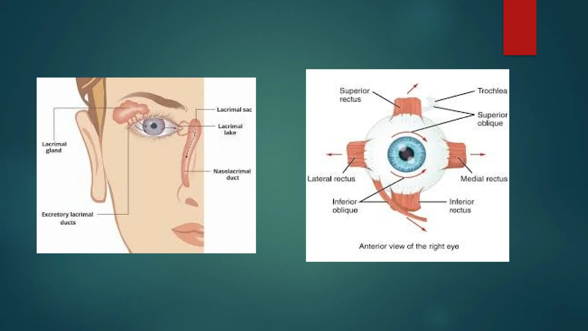
Lacrimal apparatus
Thelacrimal apertures are the small openings on the eyelids through which
tears drain from the eye into the lacrimal drainage.
Location- there are two lacrimal apertures- puncta
Upper lacrimal punctum – on the upper eyelid
Lower lacrimal punctum – on the lower eyelid
Both are located at the medial canthus(near the nose) specifically on a small
elevation called the lacrimal papilla.
Appearance & size
Tiny pin point holes, diameter is about 0.2-0.3 mm
29.
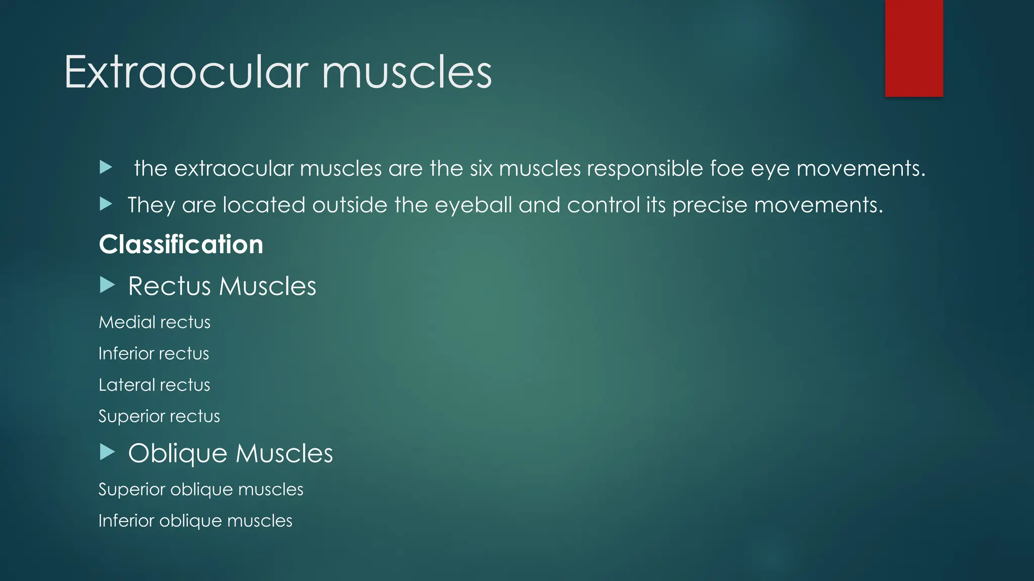
Extraocular muscles
theextraocular muscles are the six muscles responsible foe eye movements.
They are located outside the eyeball and control its precise movements.
Classification
Rectus Muscles
Medial rectus
Inferior rectus
Lateral rectus
Superior rectus
Oblique Muscles
Superior oblique muscles
Inferior oblique muscles


![Sence [vision]](https://cdn.slidesharecdn.com/ss_thumbnails/sencevision-130206202012-phpapp02-thumbnail.jpg?width=640&height=640&fit=bounds)


