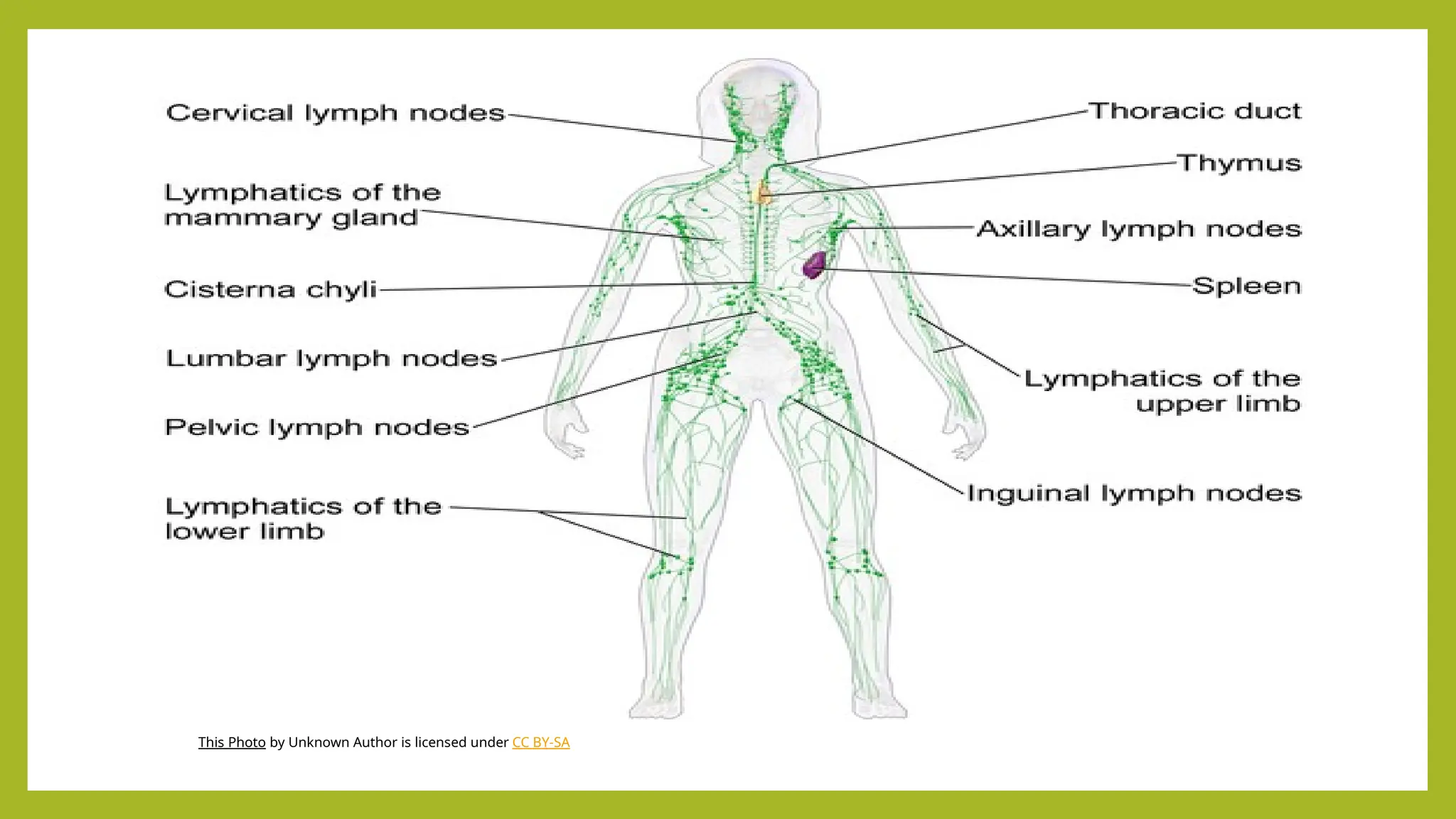
Introduction
• The lymphaticsystem is a circulatory system made up of lymph vessels, which
are much like blood vessels. It drains extra fluid (called lymph) that has passed
out of the blood and into tissues and returns it back to the blood.
• It is important part of immune system comprising a network of lymphatic
vessels that carry a clear fluid called lymph (from latin “lympha” meaning
water) directionally towards the heart.
• The tissue fluid is composed of dissolved constituents of blood and waste
materials from cells.
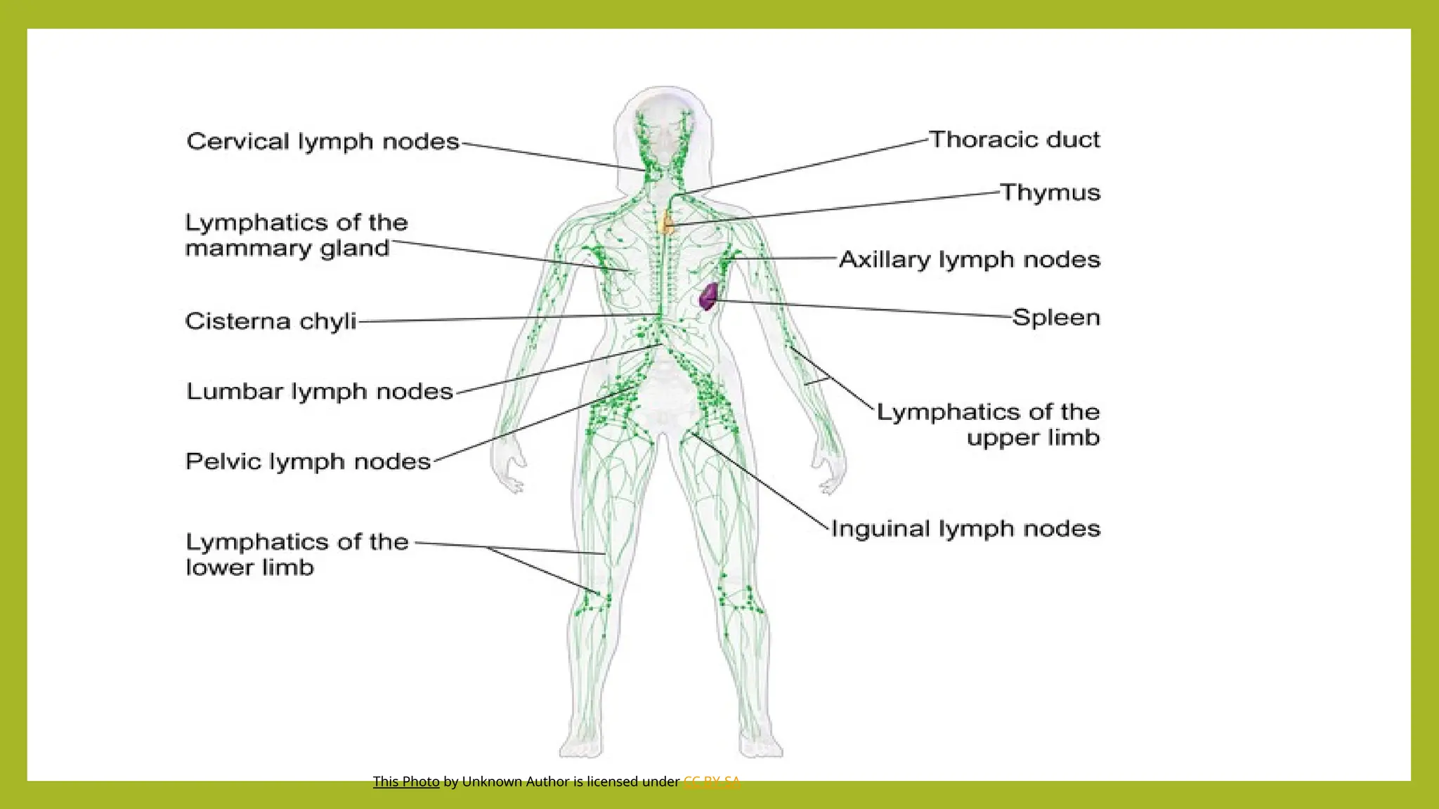
• Lymphatic system consists of lymph, lymph vessels, lymph nodes, lymph
organs, lymphoids tissue and bone marrow.
• Lymphatic system consists of lymphocytes which helps in providing immunity.
3.
Main functions oflymphatic system
1. Maintaining fluid balance by returning excess fluid from
tissues to the bloodstream.
2. Absorbing fats from the small intestine.
3. Defending the body against infection as part of the
immune system. It does this by collecting and filtering waste
products, and by producing, storing, and transporting immune
cells like lymphocytes.
4.
Other functions oflymphatic system
• Fluid Drainage:
• Collects excess interstitial fluid and proteins from tissue spaces and returns them
to the bloodstream, which helps prevent tissue swelling.
• Pathogen Filtration:
• Filters lymph through lymph nodes, which trap and destroy pathogens like
bacteria, viruses, and foreign particles.
• Protein Return:
• Returns proteins that have leaked from capillaries back into the bloodstream.
• Waste Removal:
• Collects and transports metabolic wastes, toxins, and other unwanted substances
from the tissues.
5.
.
• Fluid Homeostasis:
•Helps maintain the balance of fluid between blood and tissues.
• Nutrient and Hormone Delivery:
• Delivers nutrients and hormones to cells and tissues that are not directly supplied
by the blood capillaries.
• Cell Transport:
• Transports white blood cells and antigen-presenting cells (like dendritic cells) to the
lymph nodes to initiate an immune response.
• Abnormal Cell Removal:
• Helps remove abnormal cells, such as cancer cells, from the body's tissues and
transports them to lymph nodes for destruction.
6.
This Photo byUnknown Author is licensed under CC BY-SA
7.
Components of lymphaticsystem
1) Lymph
2) Lymph capillaries
3) Lymphatic vessels
4) Lymphoid organ
5) Lymph nodes
6) spleen
7) thymus
8) Epithelio – lymphoid system
9) Bone marrow
8.
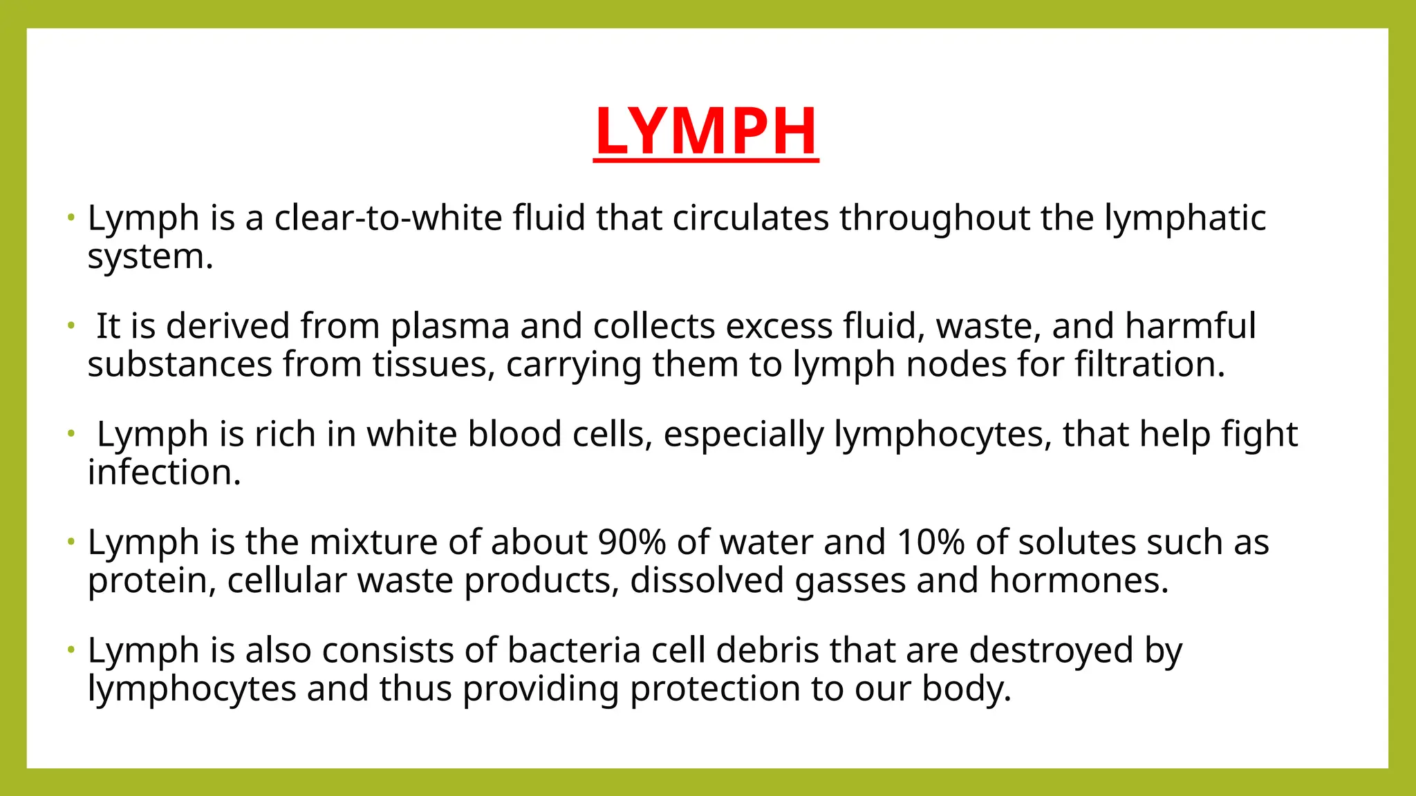
LYMPH
• Lymph isa clear-to-white fluid that circulates throughout the lymphatic
system.
• It is derived from plasma and collects excess fluid, waste, and harmful
substances from tissues, carrying them to lymph nodes for filtration.
• Lymph is rich in white blood cells, especially lymphocytes, that help fight
infection.
• Lymph is the mixture of about 90% of water and 10% of solutes such as
protein, cellular waste products, dissolved gasses and hormones.
• Lymph is also consists of bacteria cell debris that are destroyed by
lymphocytes and thus providing protection to our body.
9.
This Photo byUnknown Author is licensed under CC BY-SA
10.
Functions of thelymph
• maintaining fluid balance by returning excess tissue fluid to the
bloodstream,
• supporting the immune system by filtering and transporting pathogens
to lymph nodes for destruction,
• facilitating the absorption of fats from the digestive system.
• Lymph also helps transport nutrients to cells and removes metabolic
waste.
11.
Lymph vessels
• Lymphvessels consists of lymph capillaries, larger lymph vessels, thoracic duct and
right lymphatic duct.
• Lymph capillaries are one end closed tiny tubes originating from the interstitial spaces.
• Lymph capillaries have the same structure as that of the blood capillaries, formed of a
single layer of endothelial cells.
• The wall of lymph capillary is highly permeable to all interstitial fluid constituents.
• The lymph capillaries join together to form larger lymph vessels.
• Larger lymph vessels have the same structure and thickness as that of the wall of
small veins.
• It has cup shaped valve allowing lymph to flow only in one direction.
• Its walls have intrinsic ability to contract and relax and its property is called lymphatic
pump.
12.
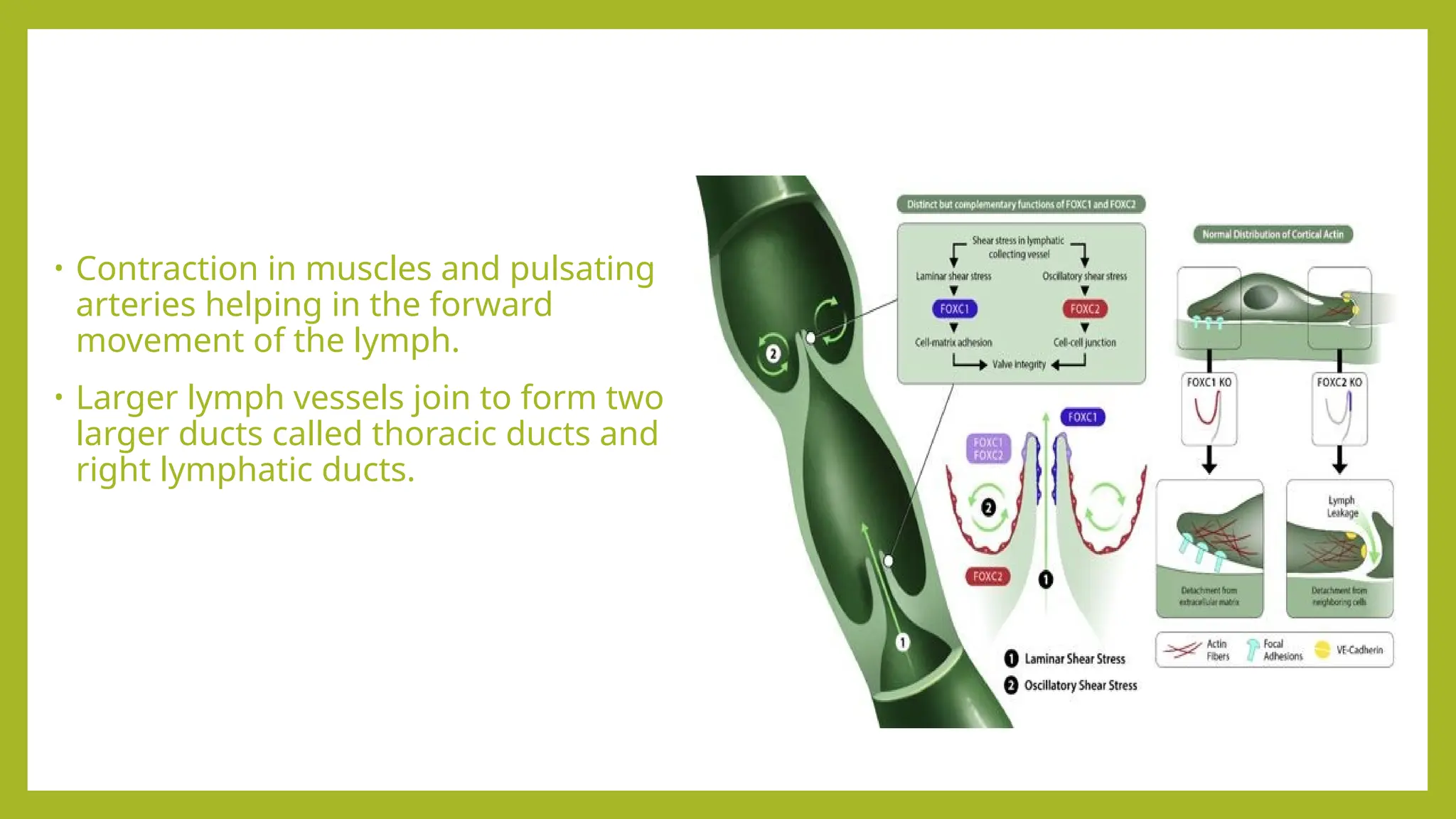
• Contraction inmuscles and pulsating
arteries helping in the forward
movement of the lymph.
• Larger lymph vessels join to form two
larger ducts called thoracic ducts and
right lymphatic ducts.
13.
Functions of lymphvessels
• Lymph vessels collect excess fluid (lymph) from tissues,
• filter it in lymph nodes, and return it to the bloodstream, which helps
maintain fluid balance, absorbs fats from the digestive system,
• aids in immune defense by transporting pathogens to the lymph nodes
for filtration.
• They act as a drainage system for tissues and play a crucial role in the
body's immune response and fat absorption.
14.
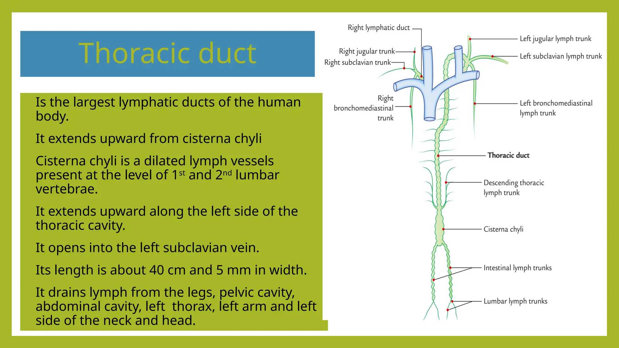
Thoracic duct
• Isthe largest lymphatic ducts of the human
body.
• It extends upward from cisterna chyli
• Cisterna chyli is a dilated lymph vessels
present at the level of 1st
and 2nd
lumbar
vertebrae.
• It extends upward along the left side of the
thoracic cavity.
• It opens into the left subclavian vein.
• Its length is about 40 cm and 5 mm in width.
• It drains lymph from the legs, pelvic cavity,
abdominal cavity, left thorax, left arm and left
side of the neck and head.
15.
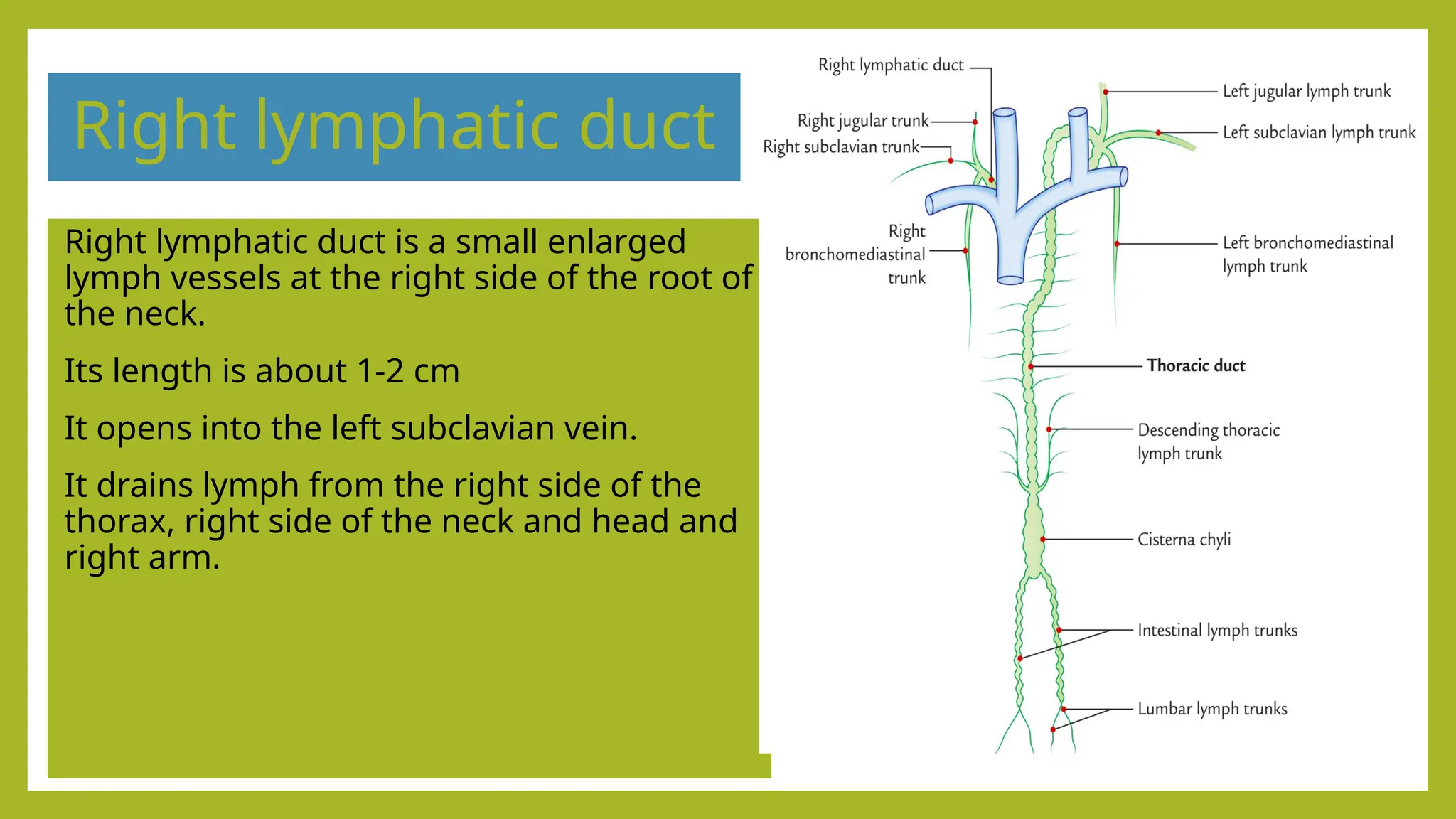
Right lymphatic duct
Rightlymphatic duct is a small enlarged
lymph vessels at the right side of the root of
the neck.
Its length is about 1-2 cm
It opens into the left subclavian vein.
It drains lymph from the right side of the
thorax, right side of the neck and head and
right arm.
16.
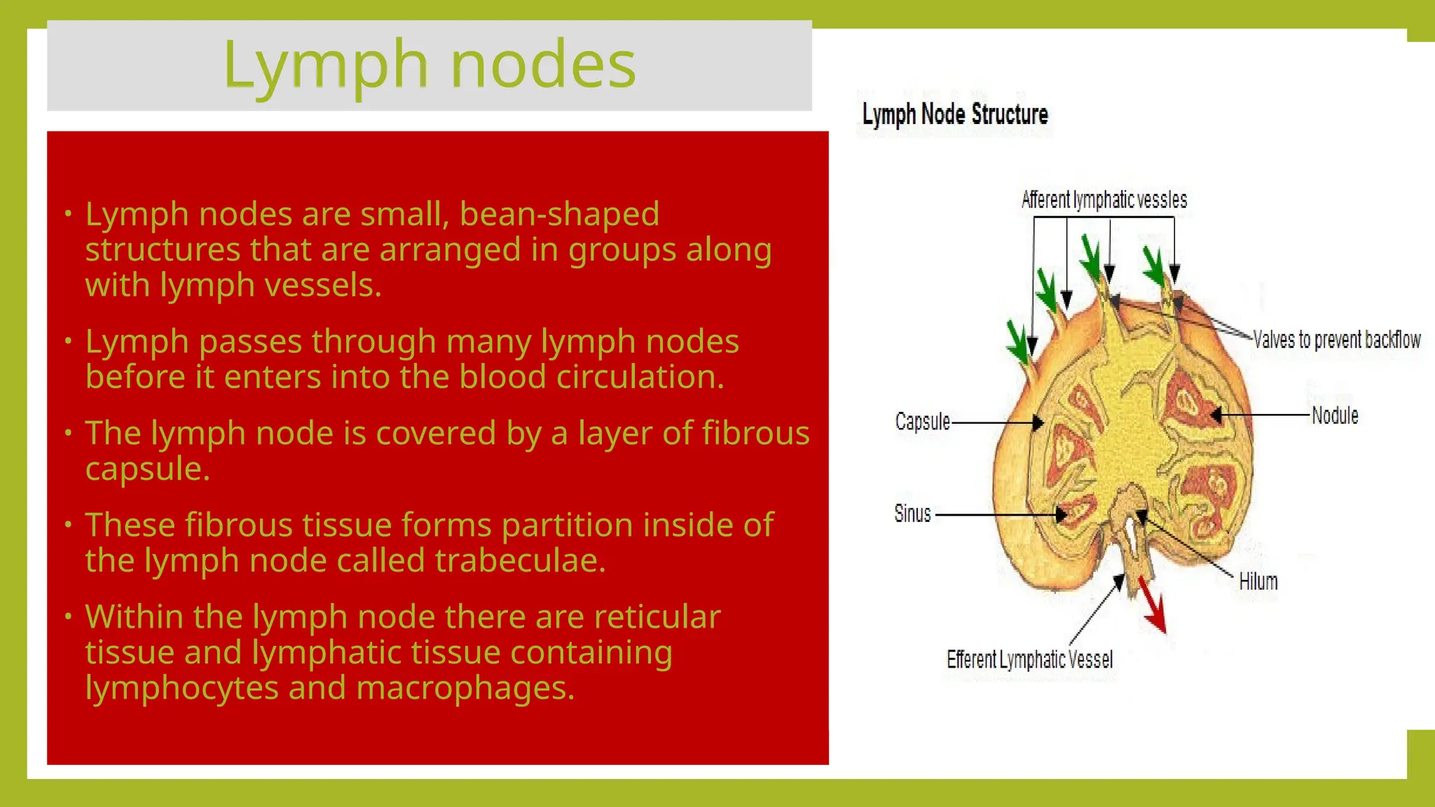
Lymph nodes
• Lymphnodes are small, bean-shaped
structures that are arranged in groups along
with lymph vessels.
• Lymph passes through many lymph nodes
before it enters into the blood circulation.
• The lymph node is covered by a layer of fibrous
capsule.
• These fibrous tissue forms partition inside of
the lymph node called trabeculae.
• Within the lymph node there are reticular
tissue and lymphatic tissue containing
lymphocytes and macrophages.
17.
Lymph nodes
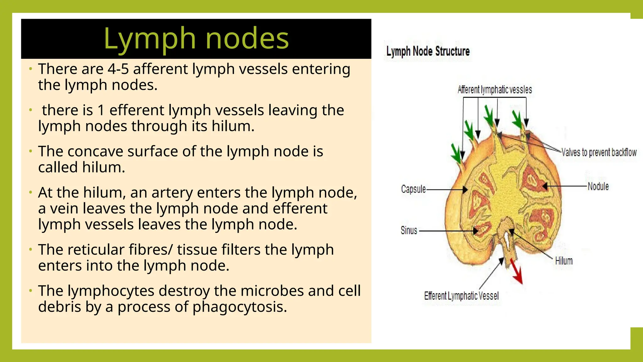
• Thereare 4-5 afferent lymph vessels entering
the lymph nodes.
• there is 1 efferent lymph vessels leaving the
lymph nodes through its hilum.
• The concave surface of the lymph node is
called hilum.
• At the hilum, an artery enters the lymph node,
a vein leaves the lymph node and efferent
lymph vessels leaves the lymph node.
• The reticular fibres/ tissue filters the lymph
enters into the lymph node.
• The lymphocytes destroy the microbes and cell
debris by a process of phagocytosis.
18.
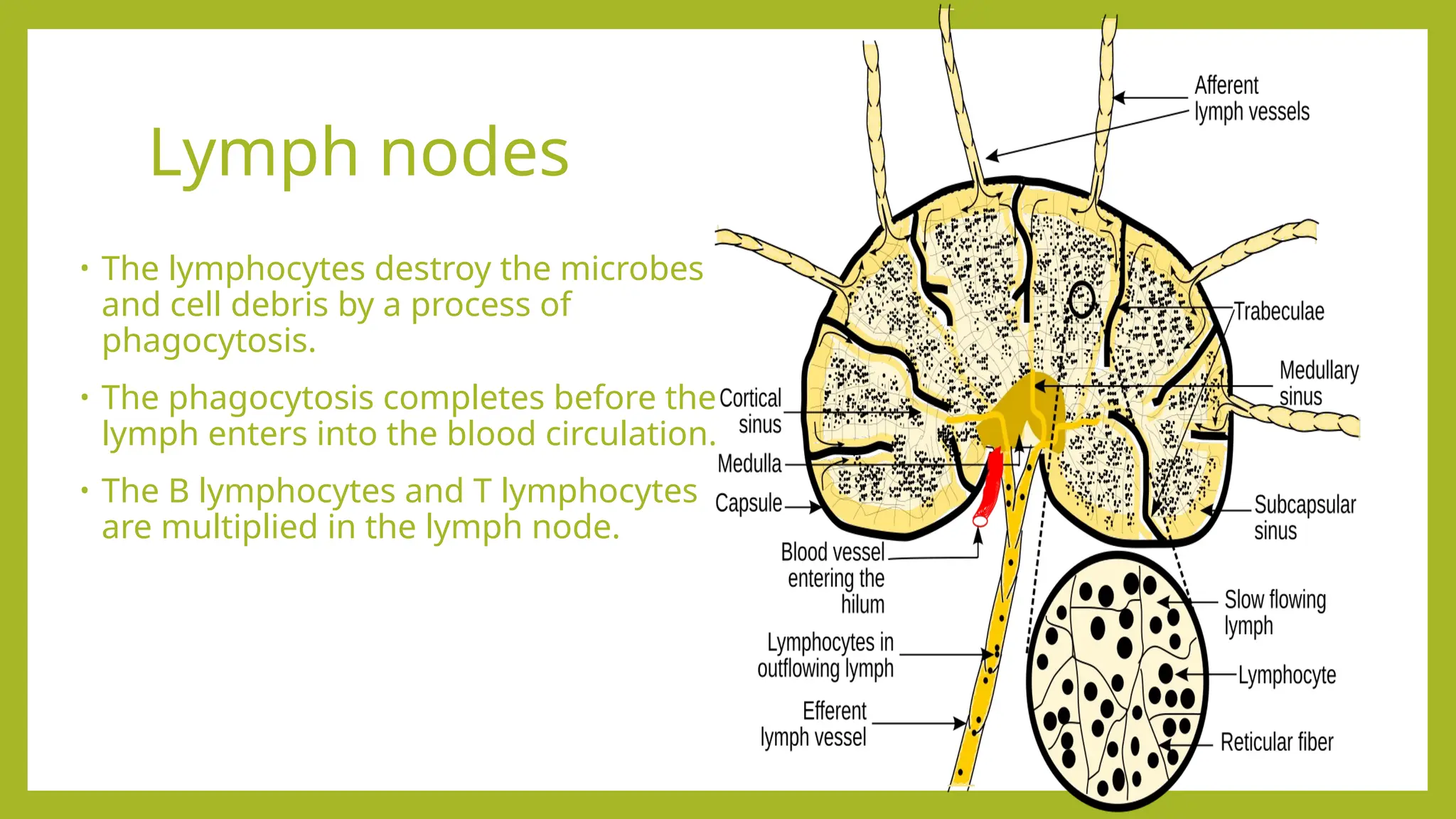
Lymph nodes
• Thelymphocytes destroy the microbes
and cell debris by a process of
phagocytosis.
• The phagocytosis completes before the
lymph enters into the blood circulation.
• The B lymphocytes and T lymphocytes
are multiplied in the lymph node.
19.
Functions of lymphnodes
•Filtering lymph fluid:
Lymph nodes filter waste, bacteria, and other foreign particles that are carried
in the lymph fluid from the body's tissues.
•Fighting infection:
They contain immune cells, primarily lymphocytes (B cells and T cells), which
identify and destroy germs, damaged cells, and abnormal cells.
•Triggering an immune response:
When a pathogen is detected, lymph nodes activate immune cells to launch an
attack against the infection, which can cause the nodes to swell.
•Returning fluid to the bloodstream:
By filtering and purifying the lymph, they help return excess fluid and proteins
back to the blood, maintaining fluid balance in the body.
20.
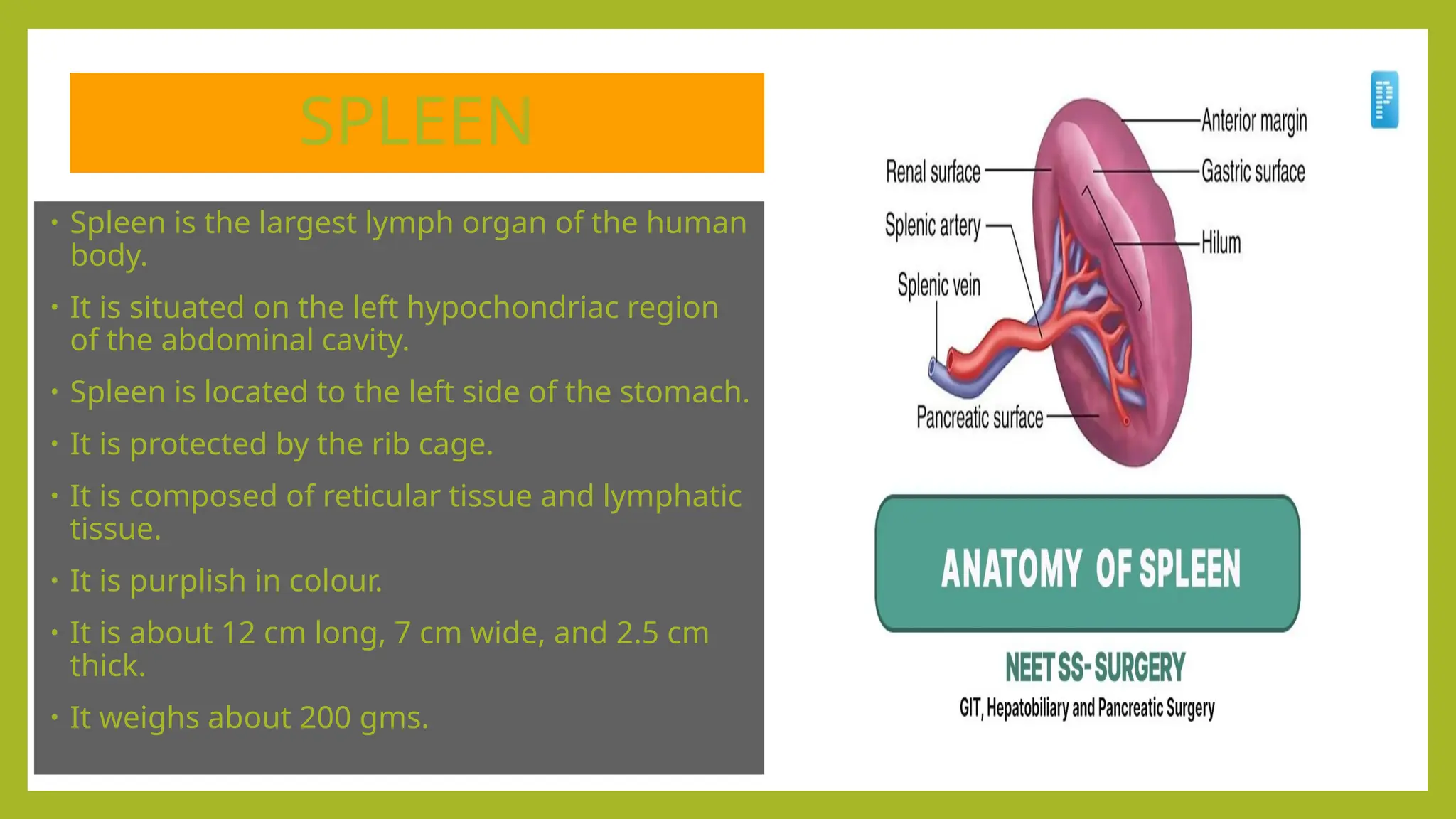
SPLEEN
• Spleen isthe largest lymph organ of the human
body.
• It is situated on the left hypochondriac region
of the abdominal cavity.
• Spleen is located to the left side of the stomach.
• It is protected by the rib cage.
• It is composed of reticular tissue and lymphatic
tissue.
• It is purplish in colour.
• It is about 12 cm long, 7 cm wide, and 2.5 cm
thick.
• It weighs about 200 gms.
21.
Spleen
• Spleen isroughly bean in shape.
• It is enclosed in a fibrous capsule.
• The structures that enter and leave the spleen through the hilum.
• The structures that enter/ leave spleen through the hilum are splenic artery, splenic vein
lymph veins and nerves.
• It acts as a filter for blood.
• Old red blood cells are recycled in the spleen.
• Old and abnormal RBCs are destroyed in spleen.
• Platelet and WBCs are stored in spleen.
• About 350 ml of blood is always stored in spleen.
• During hemorrhage, this blood is returned into the blood circulation.
• The lymphocytes protects our body from infection.
• Spleen and liver are the main sites for fetal blood cell production.
22.
Functions of Spleen
•Immunesystem support:
•Fights infection: The spleen contains white blood cells, such as lymphocytes and
macrophages, that identify and destroy germs like bacteria, viruses, and other pathogens
in the blood.
•Produces antibodies: It manufactures antibodies to help fight off infections.
•Blood filtration and maintenance:
•Filters blood: It filters blood by removing old, damaged, and abnormal red blood cells.
•Recycles iron: Macrophages in the spleen break down red blood cells and recycle their
iron.
•Stores blood cells: It stores a reserve of platelets and white blood cells, which can be
released into the bloodstream when the body needs them.
•Controls cell levels: It helps regulate the levels of red blood cells, white blood cells, and
platelets in the body.
•Emergency blood reserve:
The spleen can release stored blood into circulation in the event of significant blood loss.
•Fetal development:
During pregnancy, the spleen is partly responsible for producing hemoglobin before birth.
23.
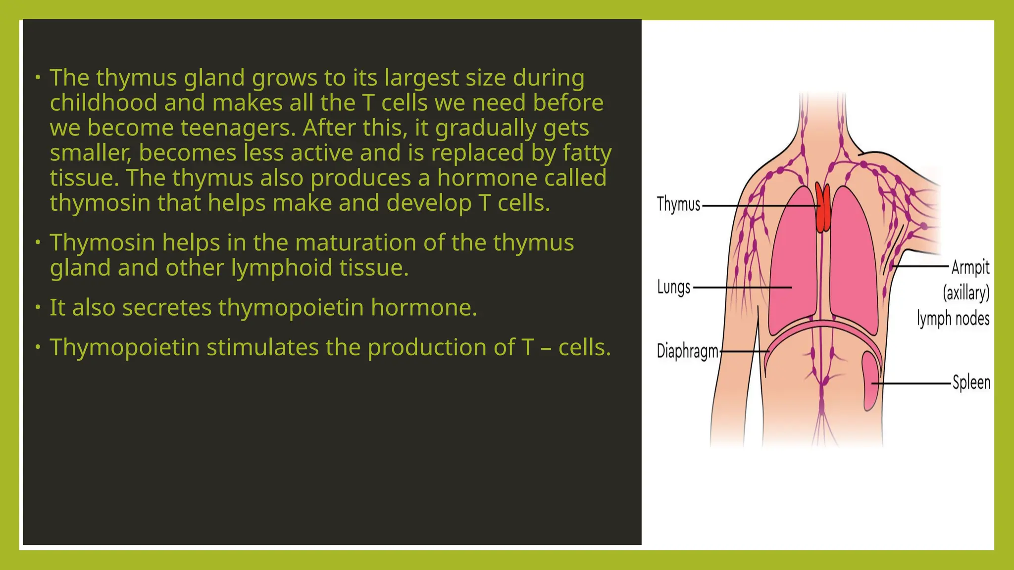
THYMUS GLAND
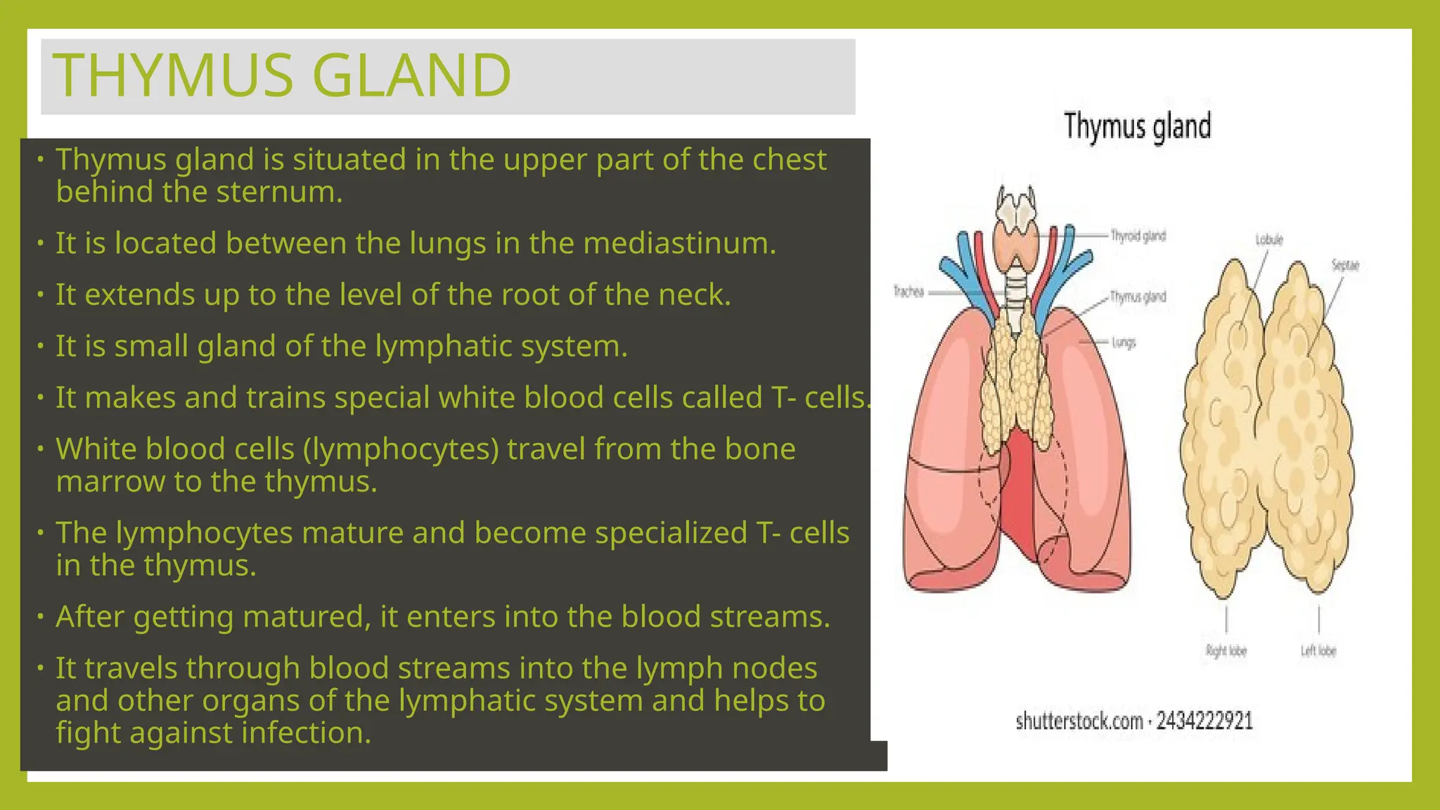
• Thymusgland is situated in the upper part of the chest
behind the sternum.
• It is located between the lungs in the mediastinum.
• It extends up to the level of the root of the neck.
• It is small gland of the lymphatic system.
• It makes and trains special white blood cells called T- cells.
• White blood cells (lymphocytes) travel from the bone
marrow to the thymus.
• The lymphocytes mature and become specialized T- cells
in the thymus.
• After getting matured, it enters into the blood streams.
• It travels through blood streams into the lymph nodes
and other organs of the lymphatic system and helps to
fight against infection.
24.
THYMUS GLAND
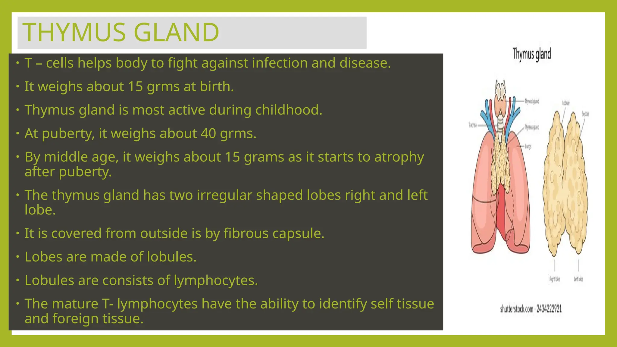
• T– cells helps body to fight against infection and disease.
• It weighs about 15 grms at birth.
• Thymus gland is most active during childhood.
• At puberty, it weighs about 40 grms.
• By middle age, it weighs about 15 grams as it starts to atrophy
after puberty.
• The thymus gland has two irregular shaped lobes right and left
lobe.
• It is covered from outside is by fibrous capsule.
• Lobes are made of lobules.
• Lobules are consists of lymphocytes.
• The mature T- lymphocytes have the ability to identify self tissue
and foreign tissue.
25.
• The thymusgland grows to its largest size during
childhood and makes all the T cells we need before
we become teenagers. After this, it gradually gets
smaller, becomes less active and is replaced by fatty
tissue. The thymus also produces a hormone called
thymosin that helps make and develop T cells.
• Thymosin helps in the maturation of the thymus
gland and other lymphoid tissue.
• It also secretes thymopoietin hormone.
• Thymopoietin stimulates the production of T – cells.
26.
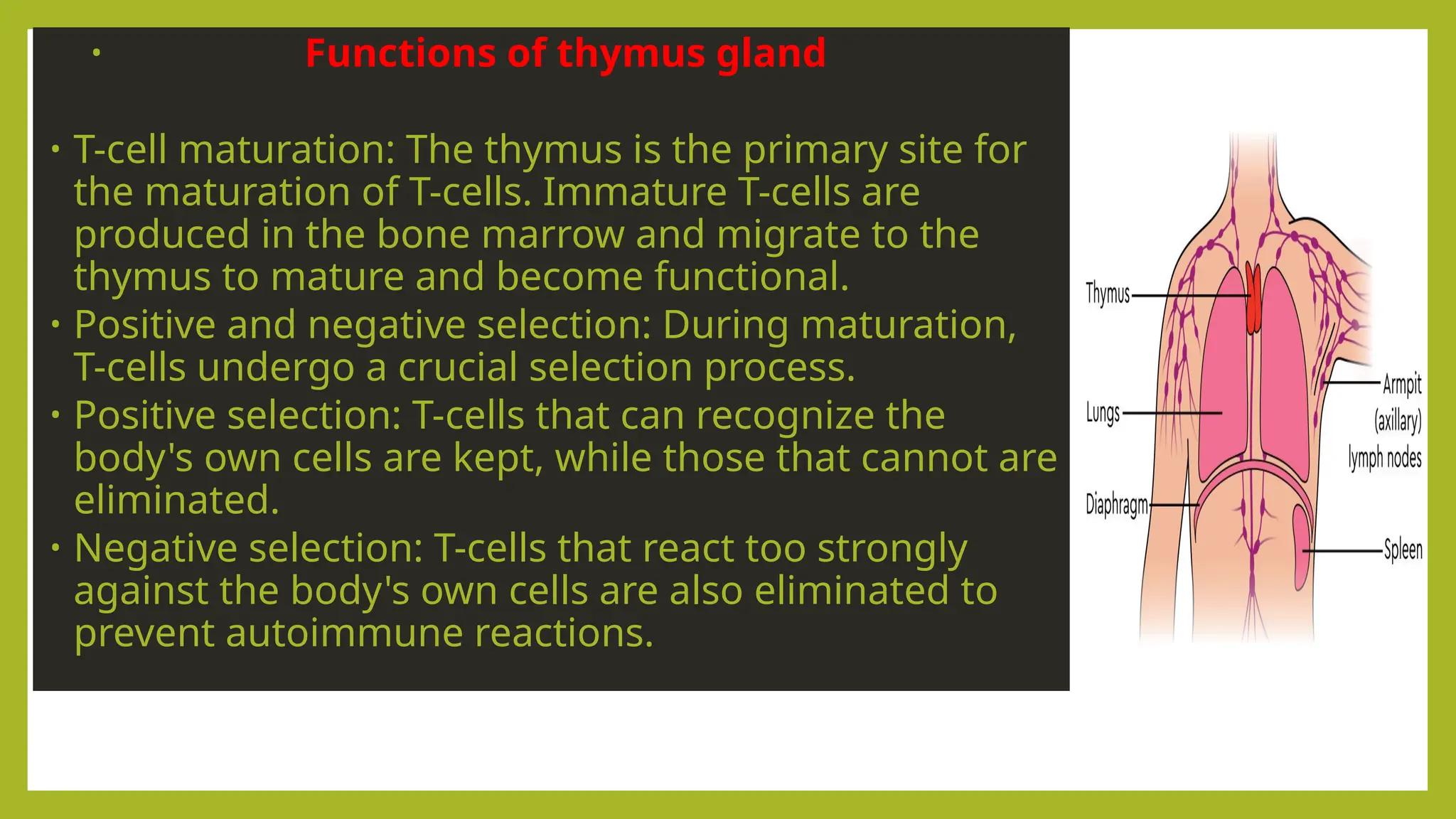
• Functions ofthymus gland
• T-cell maturation: The thymus is the primary site for
the maturation of T-cells. Immature T-cells are
produced in the bone marrow and migrate to the
thymus to mature and become functional.
• Positive and negative selection: During maturation,
T-cells undergo a crucial selection process.
• Positive selection: T-cells that can recognize the
body's own cells are kept, while those that cannot are
eliminated.
• Negative selection: T-cells that react too strongly
against the body's own cells are also eliminated to
prevent autoimmune reactions.
27.
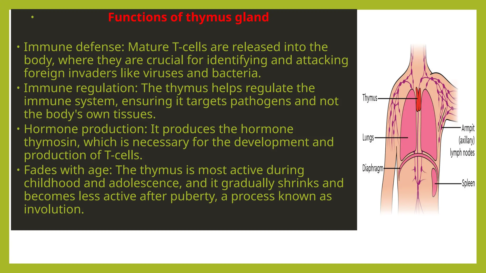
• Functions ofthymus gland
• Immune defense: Mature T-cells are released into the
body, where they are crucial for identifying and attacking
foreign invaders like viruses and bacteria.
• Immune regulation: The thymus helps regulate the
immune system, ensuring it targets pathogens and not
the body's own tissues.
• Hormone production: It produces the hormone
thymosin, which is necessary for the development and
production of T-cells.
• Fades with age: The thymus is most active during
childhood and adolescence, and it gradually shrinks and
becomes less active after puberty, a process known as
involution.
28.
MUCOSA ASSOCIATED LYMPHOID
TISSUE(MALT)
• These are collection of lymphoid tissues located at specific locations of
our body.
• These are located in the gastrointestinal tract, respiratory tract and
genitourinary tract.
• Two main types of MALT are tonsils and peyer’s patches.
•
29.
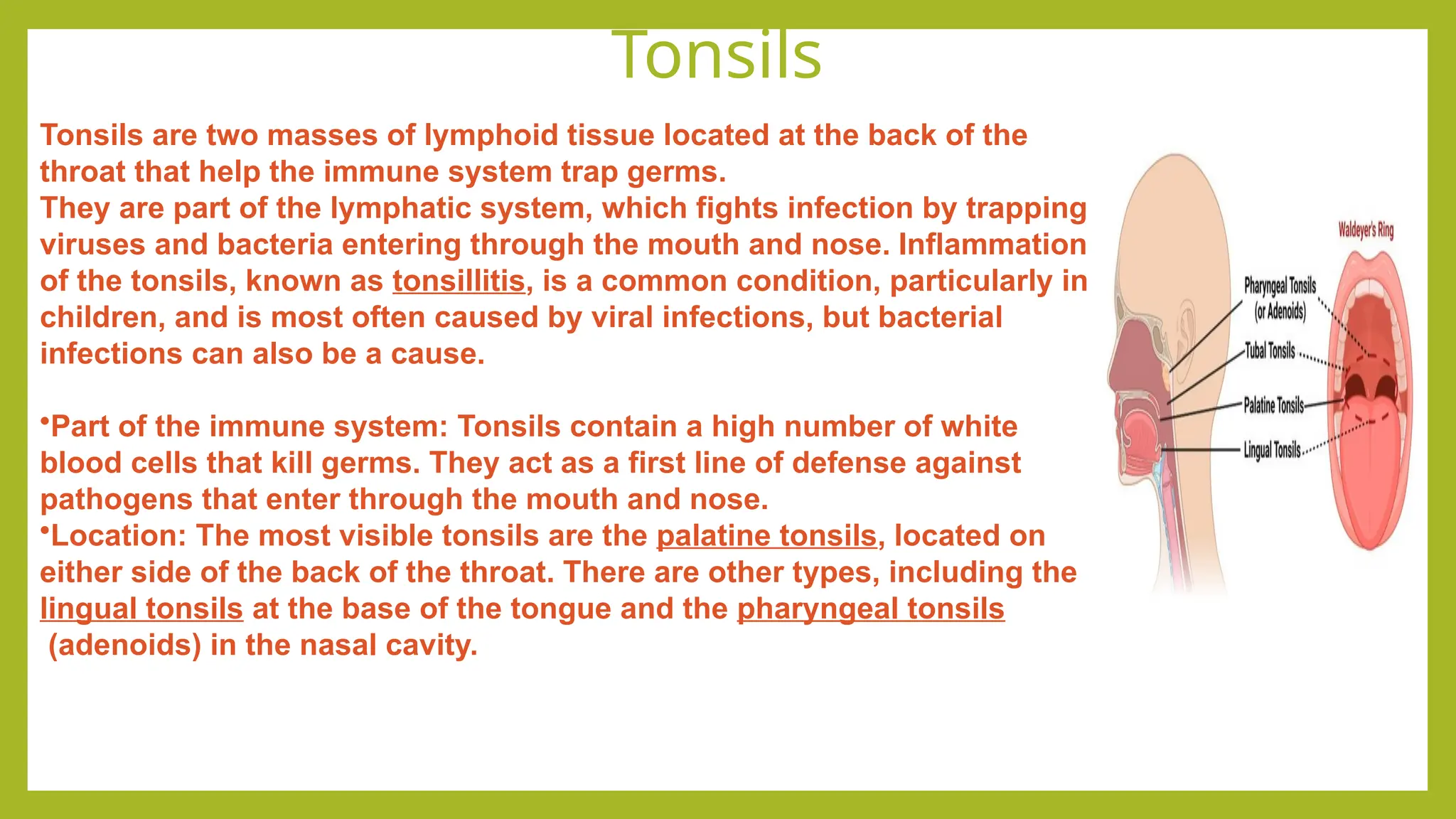
Tonsils
Tonsils are twomasses of lymphoid tissue located at the back of the
throat that help the immune system trap germs.
They are part of the lymphatic system, which fights infection by trapping
viruses and bacteria entering through the mouth and nose. Inflammation
of the tonsils, known as tonsillitis, is a common condition, particularly in
children, and is most often caused by viral infections, but bacterial
infections can also be a cause.
•Part of the immune system: Tonsils contain a high number of white
blood cells that kill germs. They act as a first line of defense against
pathogens that enter through the mouth and nose.
•Location: The most visible tonsils are the palatine tonsils, located on
either side of the back of the throat. There are other types, including the
lingual tonsils at the base of the tongue and the pharyngeal tonsils
(adenoids) in the nasal cavity.
30.
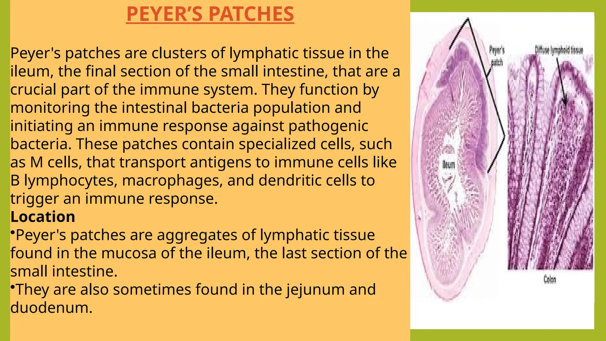
PAYER’S PATCHES
• Payer’spatches are aggregated
lymphoid follicles.
• These are situated in the inner lining
of the small intestine.
PEYER’S PATCHES
Peyer's patches are clusters of lymphatic tissue in the
ileum, the final section of the small intestine, that are a
crucial part of the immune system. They function by
monitoring the intestinal bacteria population and
initiating an immune response against pathogenic
bacteria. These patches contain specialized cells, such
as M cells, that transport antigens to immune cells like
B lymphocytes, macrophages, and dendritic cells to
trigger an immune response.
Location
•Peyer's patches are aggregates of lymphatic tissue
found in the mucosa of the ileum, the last section of the
small intestine.
•They are also sometimes found in the jejunum and
duodenum.
31.
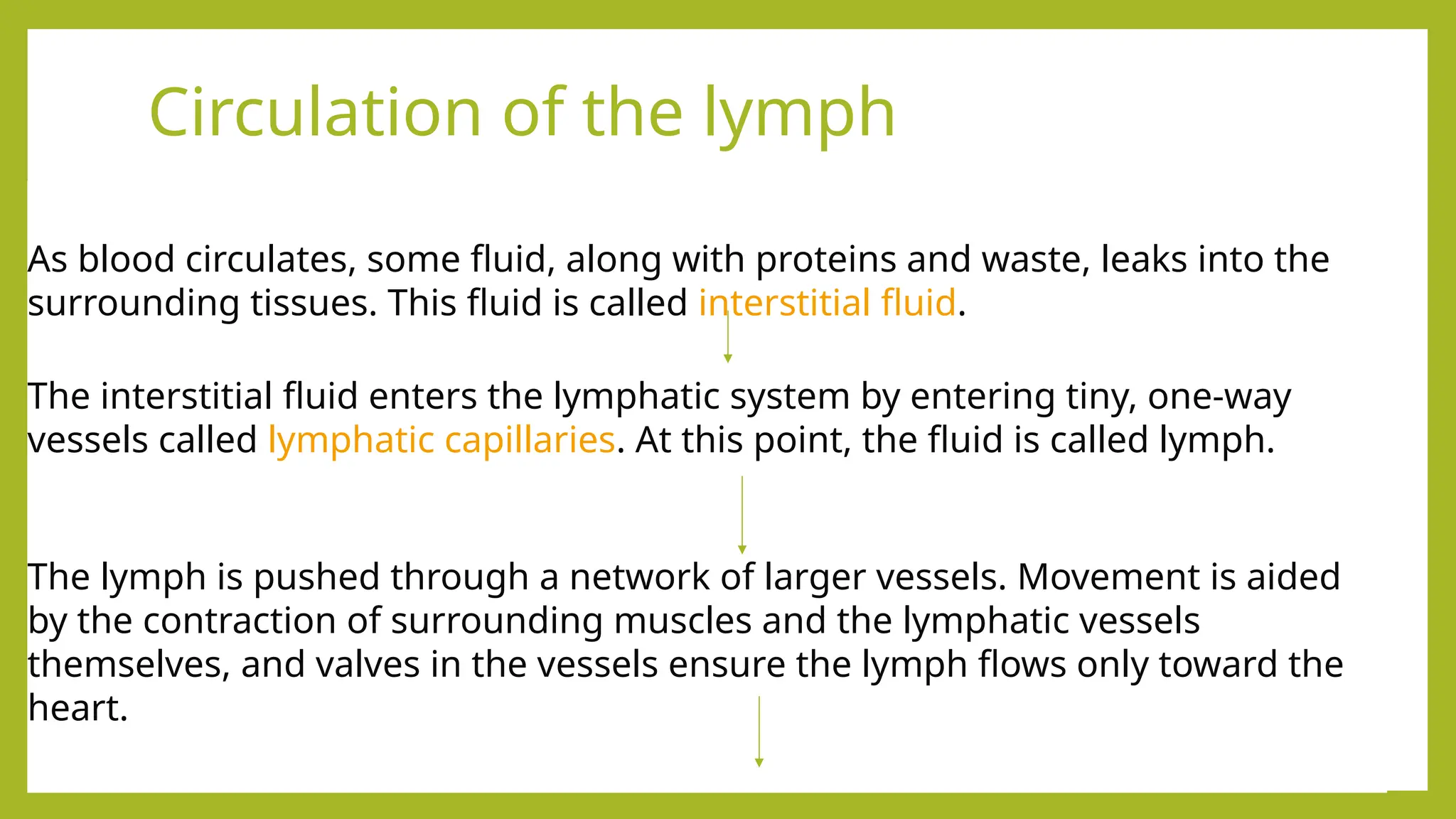
Circulation of thelymph
As blood circulates, some fluid, along with proteins and waste, leaks into the
surrounding tissues. This fluid is called interstitial fluid.
The interstitial fluid enters the lymphatic system by entering tiny, one-way
vessels called lymphatic capillaries. At this point, the fluid is called lymph.
The lymph is pushed through a network of larger vessels. Movement is aided
by the contraction of surrounding muscles and the lymphatic vessels
themselves, and valves in the vessels ensure the lymph flows only toward the
heart.
32.
Circulation of thelymph
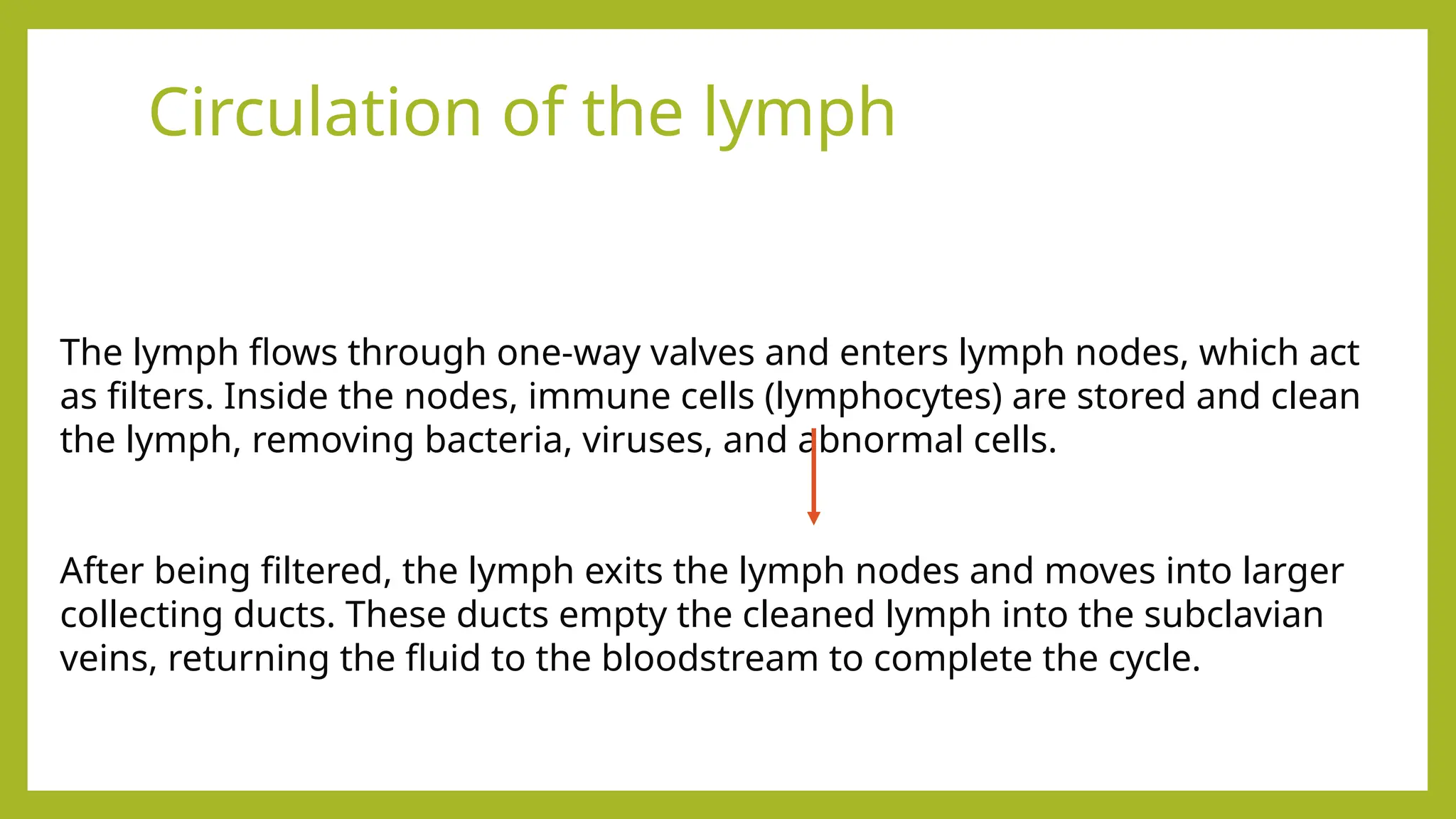
The lymph flows through one-way valves and enters lymph nodes, which act
as filters. Inside the nodes, immune cells (lymphocytes) are stored and clean
the lymph, removing bacteria, viruses, and abnormal cells.
After being filtered, the lymph exits the lymph nodes and moves into larger
collecting ducts. These ducts empty the cleaned lymph into the subclavian
veins, returning the fluid to the bloodstream to complete the cycle.
33.
Bone marrow
• Bonemarrow is a primary lymphoid organ where immune cells are
created and B cells mature. It produces all blood cells, including
lymphocytes (white blood cells), from immature stem cells. T cell
precursors are also made in the bone marrow, though they must then
travel to the thymus to fully mature.
34.
Functions of bonemarrow
•Production of immune cells: The bone marrow generates all blood cells, including the
various types of white blood cells that are central to the immune response.
•B-cell maturation: B cells are a critical component of the immune system. They are both
produced and matured in the bone marrow before they are released into the bloodstream
to circulate and seek out pathogens.
•T-cell precursor production: Bone marrow produces immature T cells, which then travel
to the thymus for further development and maturation.
•Lymphocyte production: Bone marrow is the primary site for the creation of
lymphocytes, which are a type of white blood cell that plays a key role in fighting infection.
35.
This Photo byUnknown Author is licensed under CC BY-SA


![Lymphatic system ppt[1].pptx .](https://cdn.slidesharecdn.com/ss_thumbnails/lymphaticsystemppt1-250112055350-023a9ad6-thumbnail.jpg?width=640&height=640&fit=bounds)


