Objectives for pathologylectures :
Rapid Progressive Glomerulonephritis, Chronic kidney Disease,
AND
Nephrotic and Nephritic Syndrome:
At the end of the activity) the students will be able to:
§ Recognize the five major renal glomerular syndromes.
§ Describe the main differential pathological diagnosis for each syndrome.
§ Perform a clinico-pathological correlation.
§ Describe the patterns of injury of each syndrome.
Key Outlines:
§ The nephrotic syndrome: (Minimal change, FSGS, membranous, diabetes).
§ The nephritic syndrome: (Acute post streptococcal Glomerulonephritis GN,
Membrano-proliferative GN, Systemic Lupus Erythematosus).
§ Rapidly progressive GN: (Crescentic GN)
§ Asymptomatic Hematuria / Proteinuria: IgA Nephropathy.
§ The Chronic Renal Failure.
3.
Lecture outline
§ Introduction
§Pathogenesis of glomerular disease
§ Nephrotic Syndrome
§ Minimal change disease
§ Focal segmental glomerulosclerosis
§ Membranous GN
§ Diabetes mellitus
§ Nephritic Syndrome
§ Acute post-streptococcal GN
§ Introduction to lupus nephritis
§ Introduction to membranoproliferative GN
§ Asymptomatic hematuria
§ IgA Nephropathy
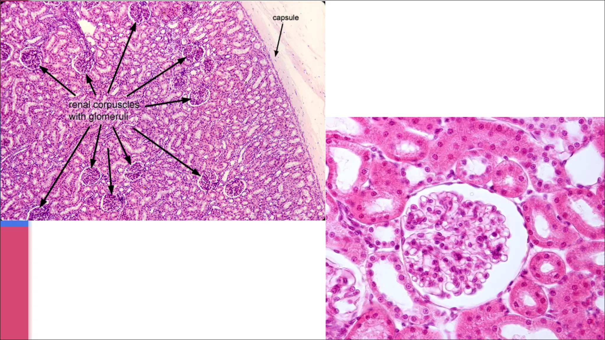
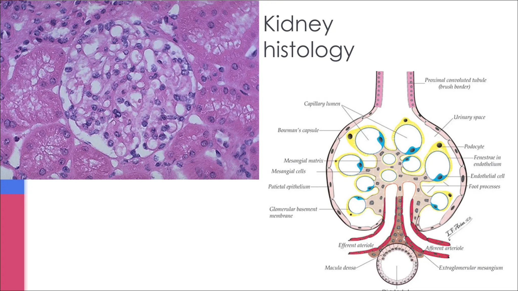
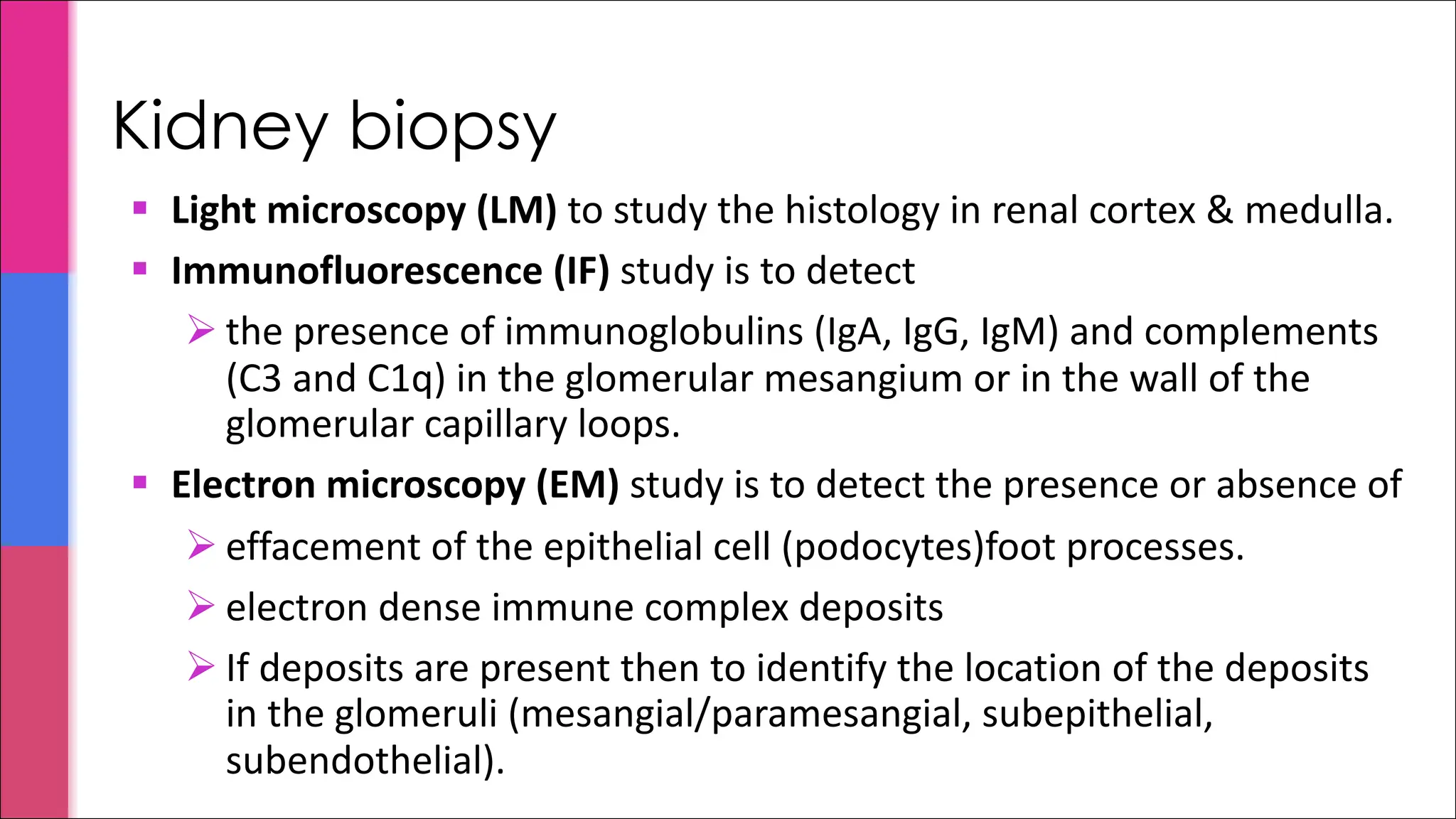
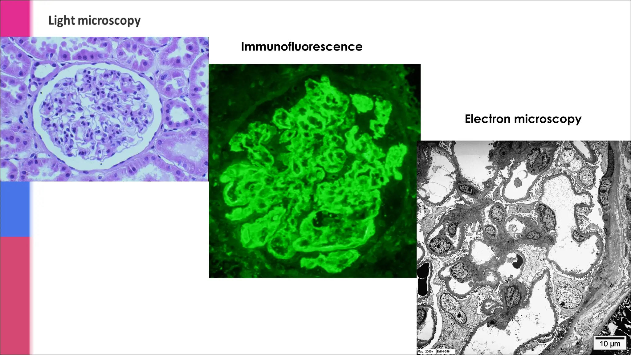
§ Light microscopy(LM) to study the histology in renal cortex & medulla.
§ Immunofluorescence (IF) study is to detect
Ø the presence of immunoglobulins (IgA, IgG, IgM) and complements
(C3 and C1q) in the glomerular mesangium or in the wall of the
glomerular capillary loops.
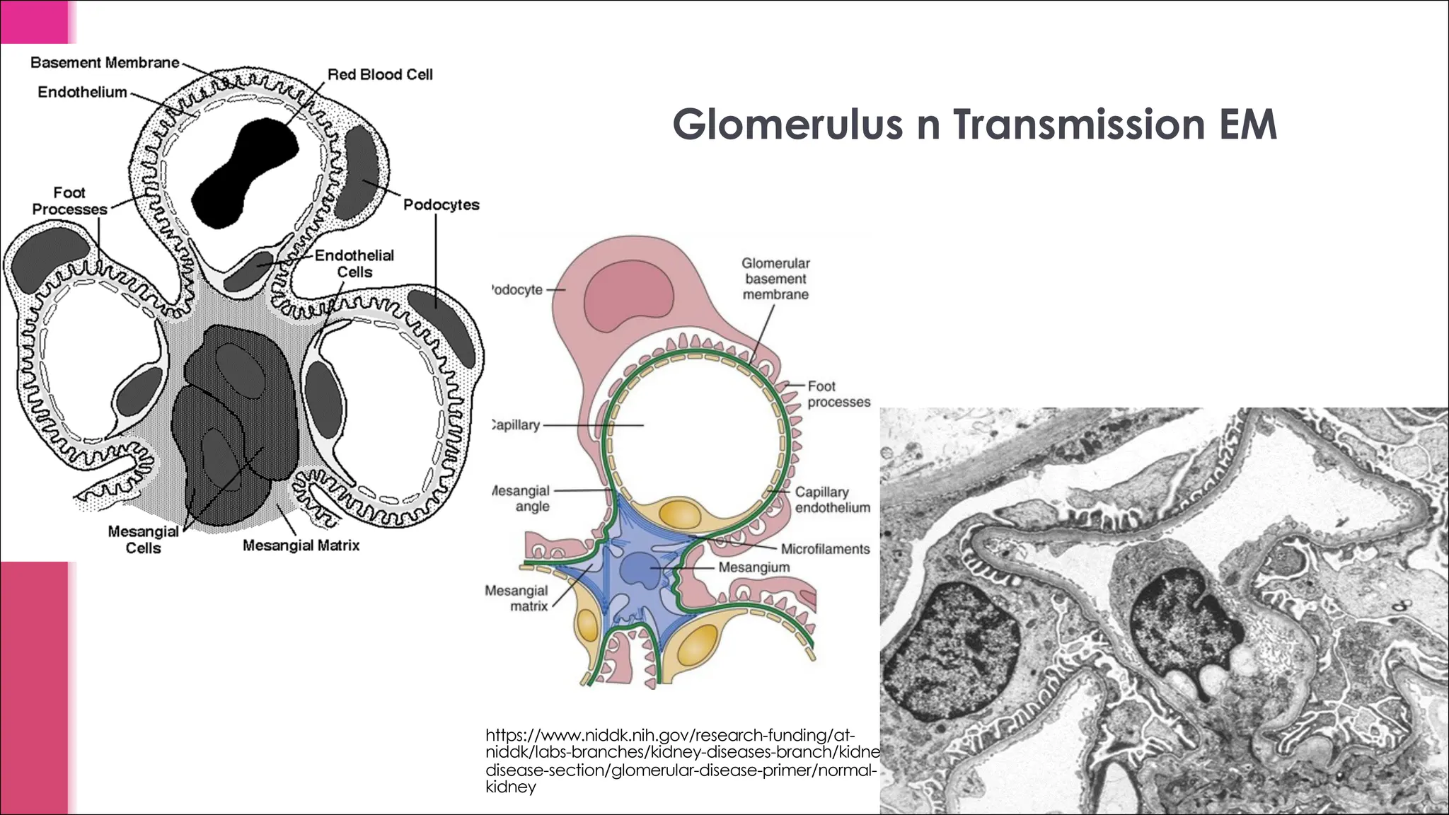
§ Electron microscopy (EM) study is to detect the presence or absence of
Ø effacement of the epithelial cell (podocytes)foot processes.
Ø electron dense immune complex deposits
Ø If deposits are present then to identify the location of the deposits
in the glomeruli (mesangial/paramesangial, subepithelial,
subendothelial).
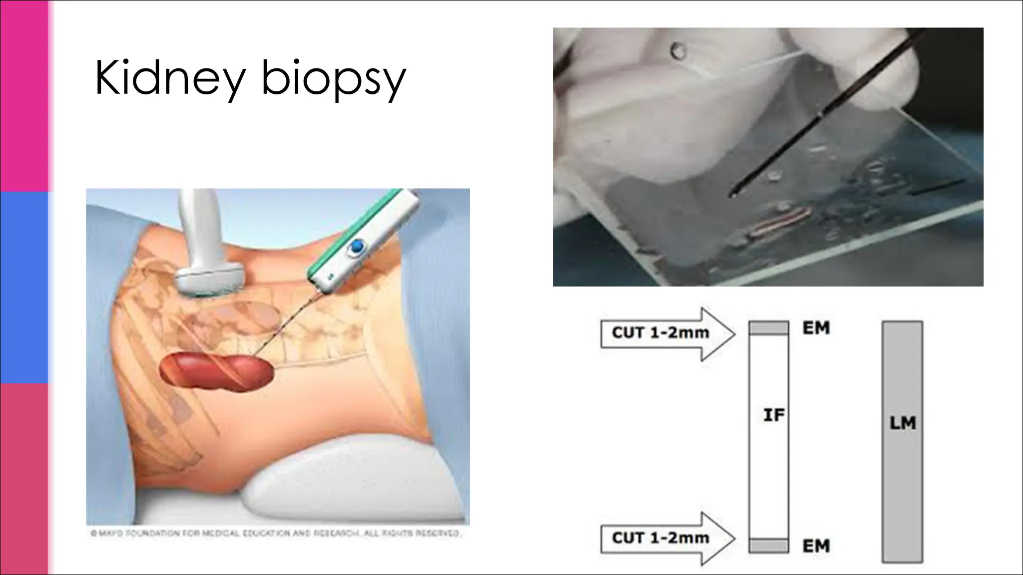
Kidney biopsy
Pathogenesis of glomerularinjury
Glomerulonephritis (GN) is frequently caused by immunologic mechanisms. Both antibody-mediated
(mainly) and cell-mediated types of immunity play roles in the production of glomerular inflammation.
1. Antibody-mediated immune GN: there are 3 major mechanisms of antibody-mediated inflammation in
most forms of GN. They are:
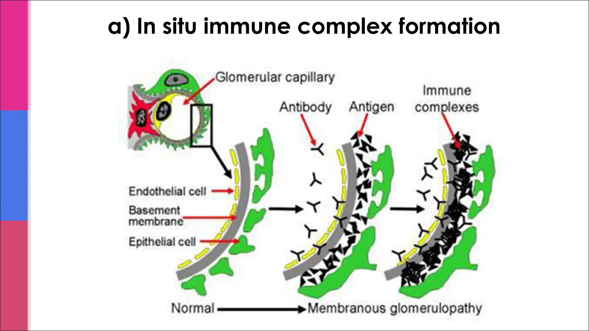
a) In situ immune complex formation: certain circulating antibodies react with certain antigens within
glomeruli à formation of immune complexes in the glomeruli. These deposits attract leukocytes and
activate complement àglomerular injury.
b) Deposition of preformed circulating immune complexes in glomeruli à deposits attract
inflammatory cells and activate complements à glomerular injury (e.g. antigens released by
bacteria or virus can bind to circulating antibodies to produce immune complexes à can deposit in
glomeruli).
c) Antineutrophil cytoplasmic autoantibodies (ANCAs): cause a severe GN. Patients have circulating
autoantibodies against antigens in the cytoplasm of neutrophils. This interaction leads to activation
and adhesion of the neutrophils to endothelial cells lining the capillaries especially the glomerular
capillaries. The neutrophils release injurious products that promote vascular inflammation and GN
All 3 initiate of glomerular inflammatory injury by attraction and activation of leukocytes.
2. Cell mediated immune GN: sensitized T cells can also cause glomerular injury.
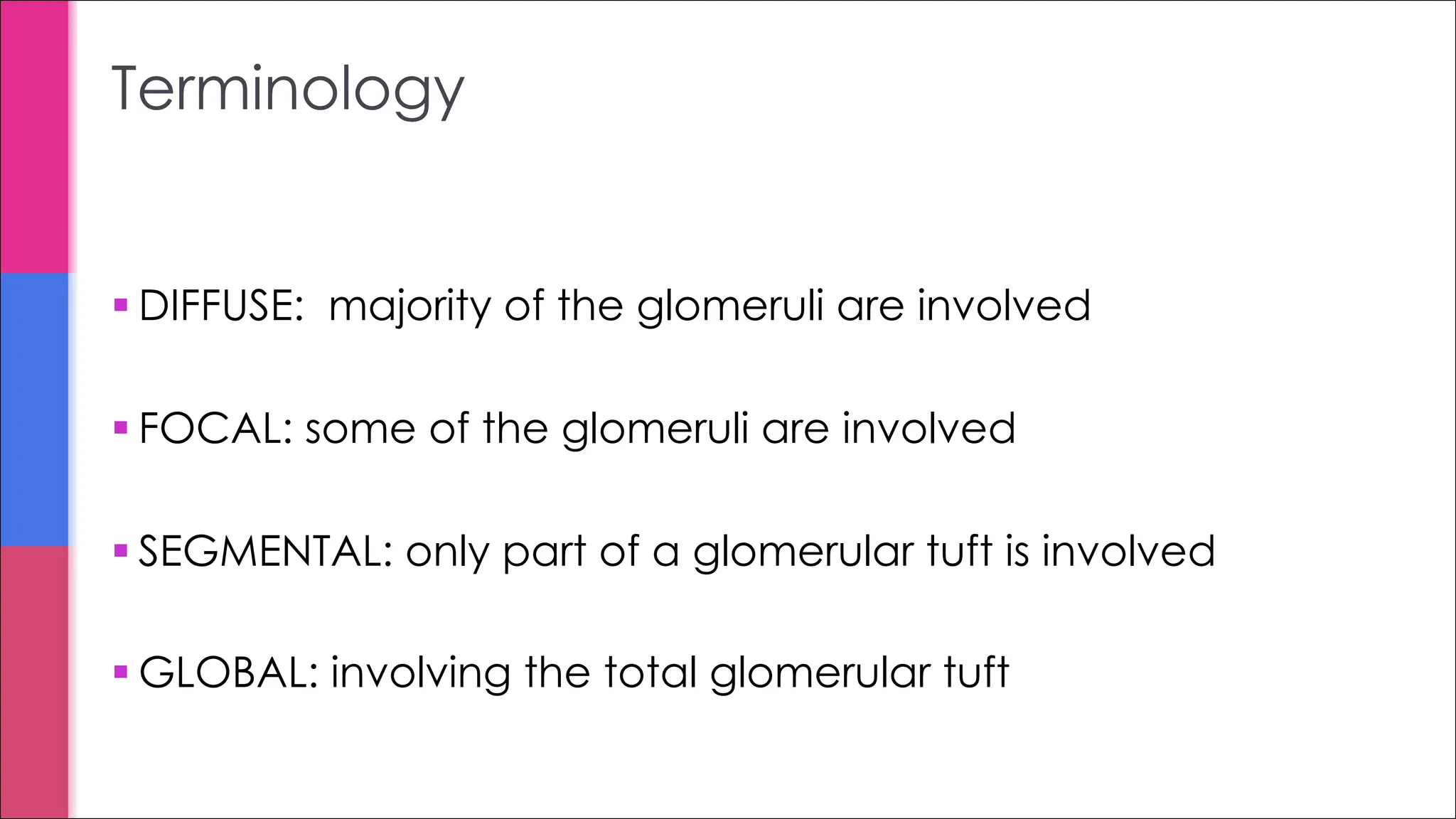
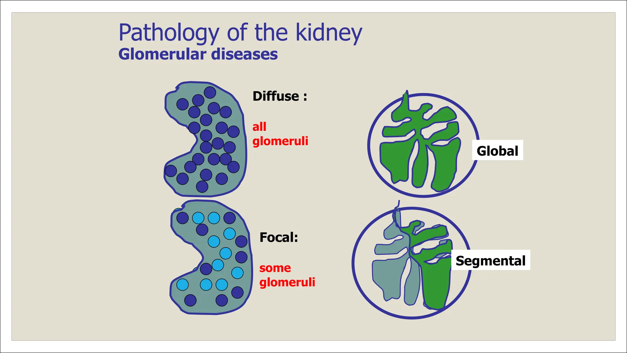
§ DIFFUSE: majorityof the glomeruli are involved
§ FOCAL: some of the glomeruli are involved
§ SEGMENTAL: only part of a glomerular tuft is involved
§ GLOBAL: involving the total glomerular tuft
Terminology
13.
Pathology of thekidney
Glomerular diseases
Diffuse :
all
glomeruli
Focal:
some
glomeruli
Global
Segmental
14.
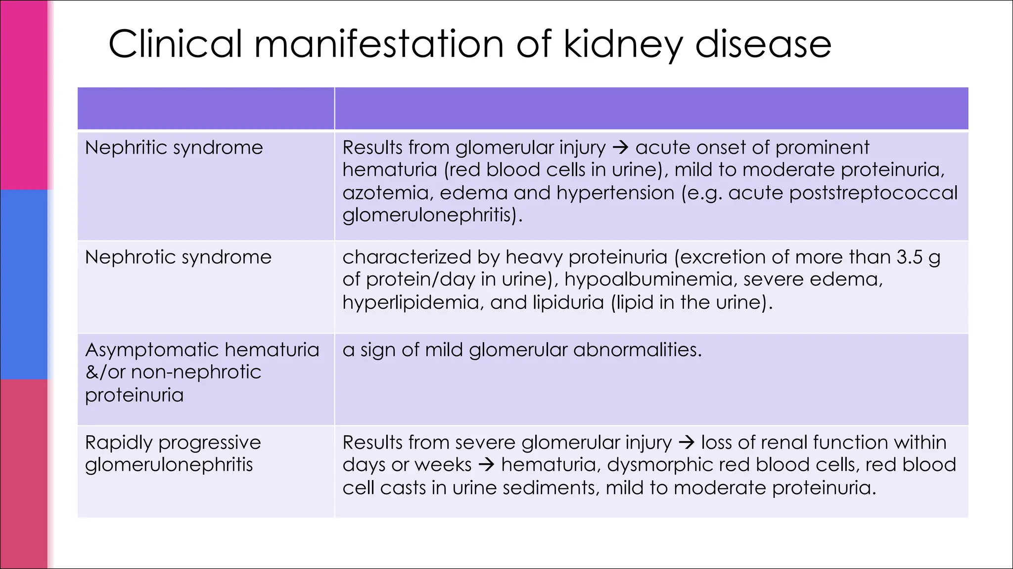
Clinical manifestation ofkidney disease
Nephritic syndrome Results from glomerular injury à acute onset of prominent
hematuria (red blood cells in urine), mild to moderate proteinuria,
azotemia, edema and hypertension (e.g. acute poststreptococcal
glomerulonephritis).
Nephrotic syndrome characterized by heavy proteinuria (excretion of more than 3.5 g
of protein/day in urine), hypoalbuminemia, severe edema,
hyperlipidemia, and lipiduria (lipid in the urine).
Asymptomatic hematuria
&/or non-nephrotic
proteinuria
a sign of mild glomerular abnormalities.
Rapidly progressive
glomerulonephritis
Results from severe glomerular injury à loss of renal function within
days or weeks à hematuria, dysmorphic red blood cells, red blood
cell casts in urine sediments, mild to moderate proteinuria.
15.
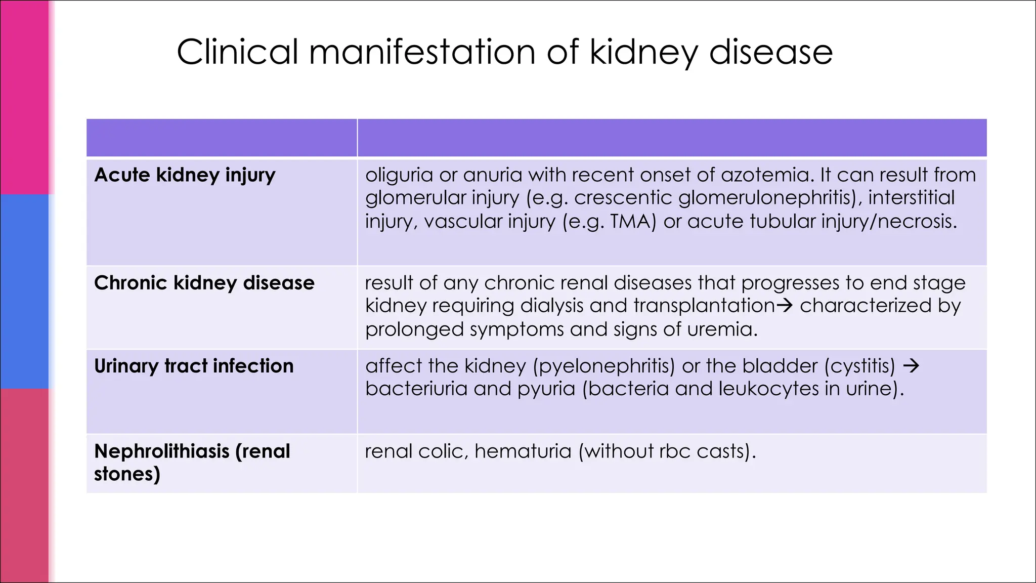
Clinical manifestation ofkidney disease
Acute kidney injury oliguria or anuria with recent onset of azotemia. It can result from
glomerular injury (e.g. crescentic glomerulonephritis), interstitial
injury, vascular injury (e.g. TMA) or acute tubular injury/necrosis.
Chronic kidney disease result of any chronic renal diseases that progresses to end stage
kidney requiring dialysis and transplantationà characterized by
prolonged symptoms and signs of uremia.
Urinary tract infection affect the kidney (pyelonephritis) or the bladder (cystitis) à
bacteriuria and pyuria (bacteria and leukocytes in urine).
Nephrolithiasis (renal
stones)
renal colic, hematuria (without rbc casts).
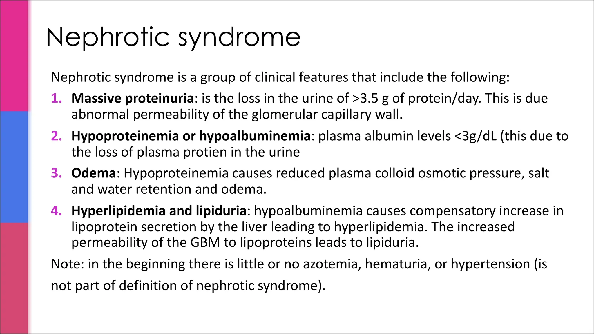
Nephrotic syndrome
Nephrotic syndromeis a group of clinical features that include the following:
1. Massive proteinuria: is the loss in the urine of >3.5 g of protein/day. This is due
abnormal permeability of the glomerular capillary wall.
2. Hypoproteinemia or hypoalbuminemia: plasma albumin levels <3g/dL (this due to
the loss of plasma protien in the urine
3. Odema: Hypoproteinemia causes reduced plasma colloid osmotic pressure, salt
and water retention and odema.
4. Hyperlipidemia and lipiduria: hypoalbuminemia causes compensatory increase in
lipoprotein secretion by the liver leading to hyperlipidemia. The increased
permeability of the GBM to lipoproteins leads to lipiduria.
Note: in the beginning there is little or no azotemia, hematuria, or hypertension (is
not part of definition of nephrotic syndrome).
PRIMARY CAUSES
§ Minimalchange disease
§ Membranous GN
§ Focal segmental glomerulosclerosis (FSGS)
§ Membranoproliferative GN (can also present as nephritic syndrome)
§ Others
SECONDARY CAUSES
§ Diabetes mellitus (most common systemic causes)
§ Amyloidosis
§ Systemic lupus erythematosus (it can also present as nephritic syndrome)
§ Drugs (gold, penicillamine, "street heroin")
§ Others
NOTE: In children the most common cause of nephrotic syndrome is minimal-change disease. In adults
the most common primary glomerular diseases that causes nephrotic syndrome are membranous
glomerulopathy in Caucasians and Asians, and FSGS is the most common etiology in American blacks.
Causes of nephrotic syndrome
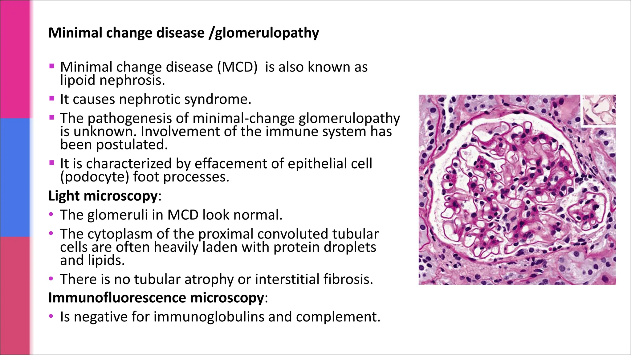
§ Minimal changedisease (MCD) is also known as
lipoid nephrosis.
§ It causes nephrotic syndrome.
§ The pathogenesis of minimal-change glomerulopathy
is unknown. Involvement of the immune system has
been postulated.
§ It is characterized by effacement of epithelial cell
(podocyte) foot processes.
Light microscopy:
• The glomeruli in MCD look normal.
• The cytoplasm of the proximal convoluted tubular
cells are often heavily laden with protein droplets
and lipids.
• There is no tubular atrophy or interstitial fibrosis.
Immunofluorescence microscopy:
• Is negative for immunoglobulins and complement.
Minimal change disease /glomerulopathy
22.
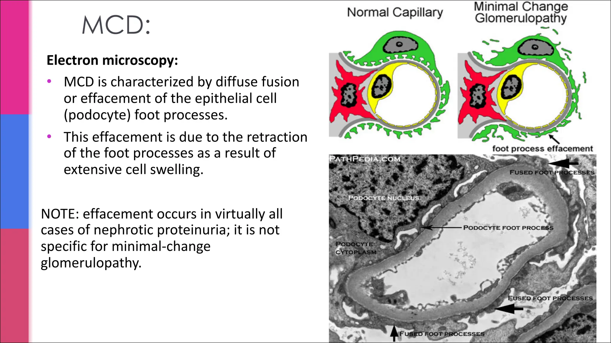
MCD:
Electron microscopy:
• MCDis characterized by diffuse fusion
or effacement of the epithelial cell
(podocyte) foot processes.
• This effacement is due to the retraction
of the foot processes as a result of
extensive cell swelling.
NOTE: effacement occurs in virtually all
cases of nephrotic proteinuria; it is not
specific for minimal-change
glomerulopathy.
23.
MCD: clinical features,treatment and prognosis
§ C/F include:
§ Nephrotic syndrome
§ Treatment and prognosis:
§ Over 90% of children and few adults have complete remission within 8 weeks
of corticosteroid therapy.
§ In the absence of complications, the prognosis of MCD, especially in children
is very good.
§ Some patients become steroid dependent i.e. after withdrawal of
corticosteroids relapses occur.
§ A small subgroup of patients has only partial remission.
§ Less than 5% develop chronic renal failure
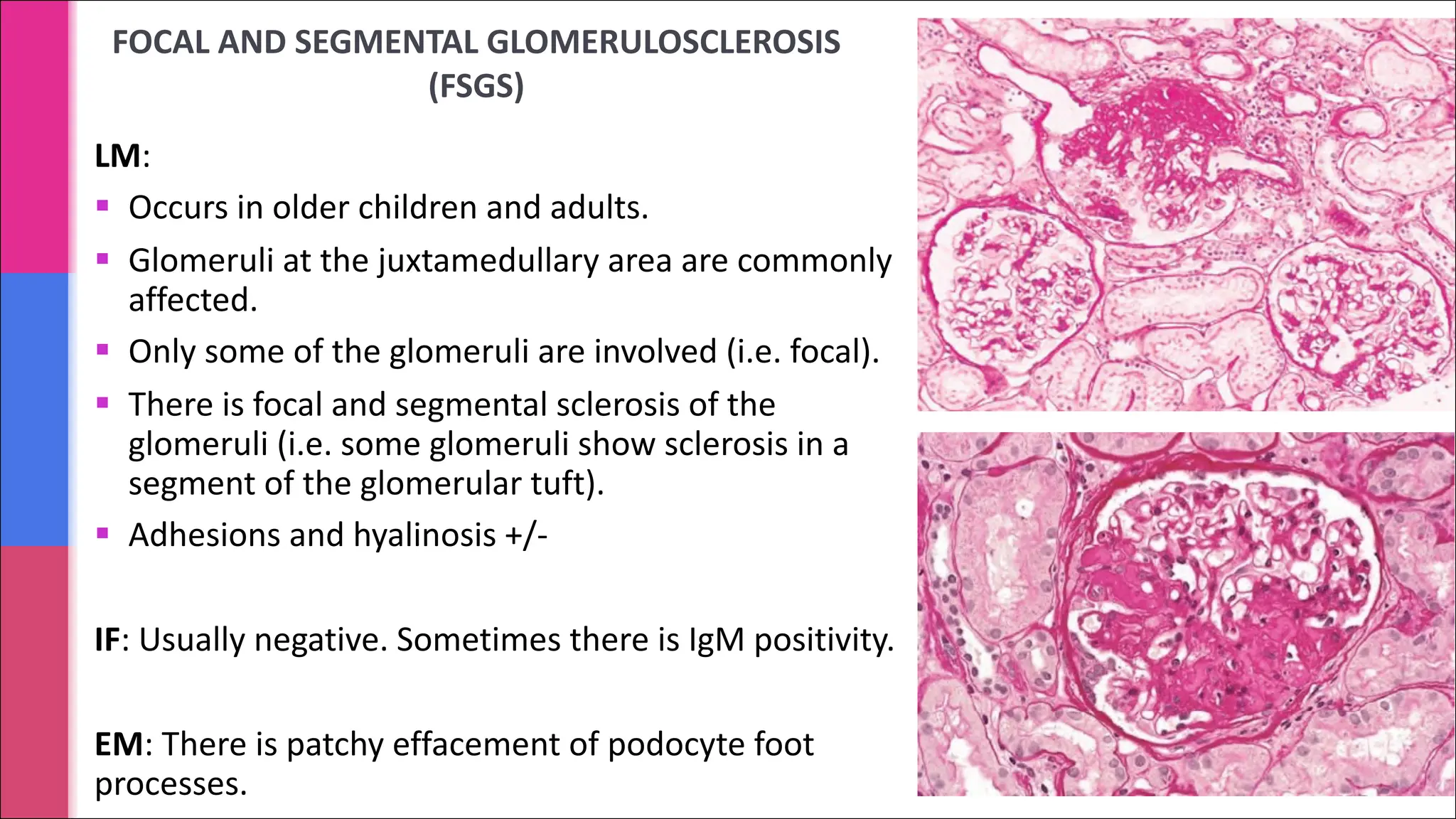
FOCAL AND SEGMENTALGLOMERULOSCLEROSIS
(FSGS)
LM:
§ Occurs in older children and adults.
§ Glomeruli at the juxtamedullary area are commonly
affected.
§ Only some of the glomeruli are involved (i.e. focal).
§ There is focal and segmental sclerosis of the
glomeruli (i.e. some glomeruli show sclerosis in a
segment of the glomerular tuft).
§ Adhesions and hyalinosis +/-
IF: Usually negative. Sometimes there is IgM positivity.
EM: There is patchy effacement of podocyte foot
processes.
§ It isa frequent cause of the nephrotic syndrome in adults (commonly 30 to 50 years)
§ It is an immune complex disease. The antigen-antibody immune complexes are formed
either in situ in the glomeruli or are preformed in circulation and then deposited in the
glomeruli.
§ It is characterized by accumulation of immune complexes in the subepithelial area in the
glomeruli (between the podocytes and the GBM).
§ It is a slowly progressive disease. If not treated it à fibrosis of the kidneys (glomerular
sclerosis, atrophy of tubules and interstitial fibrosis) and end stage disease.
§ Membranous glomerulopathy can be:
ØPrimary/idiopathic membranous GN: about 85% of cases.
ØSecondary membranous GN: causes include autoimmune disease (SLE), infectious
disease (hepatitis B), therapeutic agents (penicillamine), neoplasms (lung cancer).
Patient should be investigated for secondary causes.
Membranous glomerulopathy/ glomerulonephritis (GN)
28.
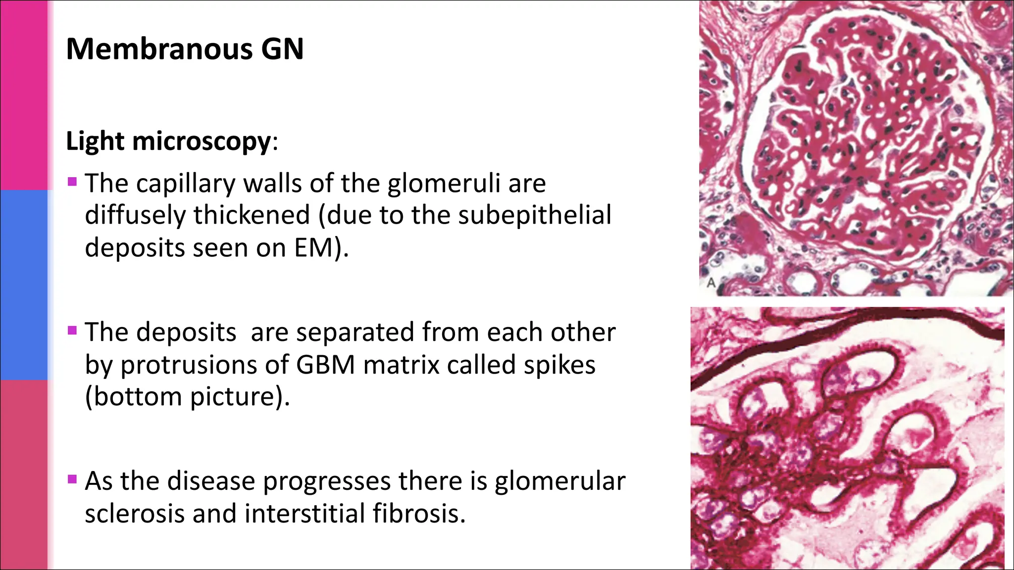
Membranous GN
Light microscopy:
§The capillary walls of the glomeruli are
diffusely thickened (due to the subepithelial
deposits seen on EM).
§ The deposits are separated from each other
by protrusions of GBM matrix called spikes
(bottom picture).
§ As the disease progresses there is glomerular
sclerosis and interstitial fibrosis.
29.
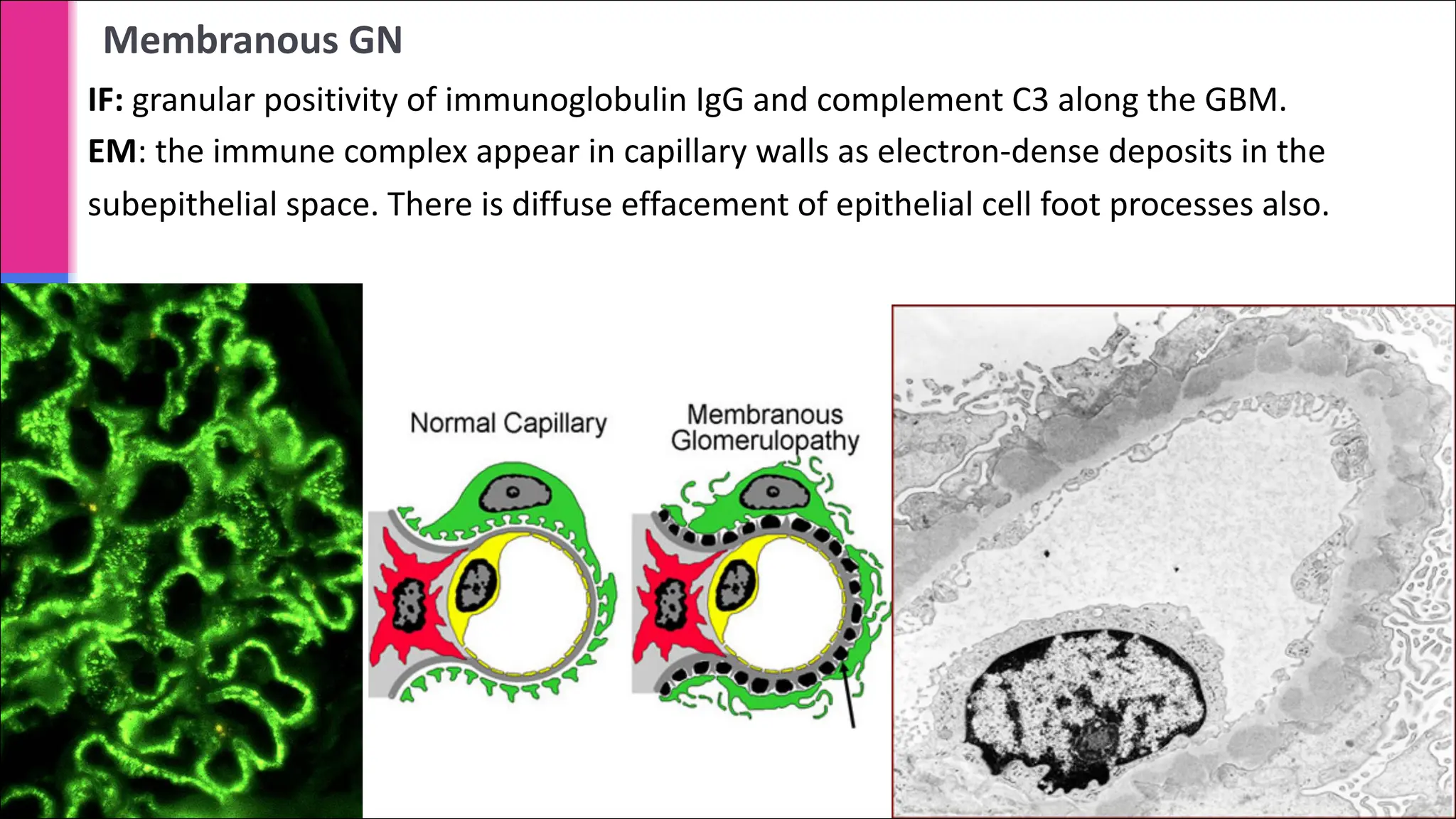
Membranous GN
IF: granularpositivity of immunoglobulin IgG and complement C3 along the GBM.
EM: the immune complex appear in capillary walls as electron-dense deposits in the
subepithelial space. There is diffuse effacement of epithelial cell foot processes also.
30.
Membranous GN/ glomerulopathy:clinical feature
§ Commonly 30 to 50 years of age
§ Nephrotic syndrome
§ The proteinuria does not usually respond to corticosteroid therapy
§ Proteinuria persists in about half the patients
§ Some case progress to renal failure
§ 10% to 30% have a more benign course with good prognosis.
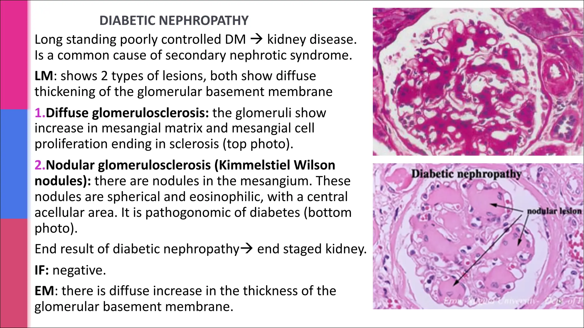
DIABETIC NEPHROPATHY
Long standingpoorly controlled DM à kidney disease.
Is a common cause of secondary nephrotic syndrome.
LM: shows 2 types of lesions, both show diffuse
thickening of the glomerular basement membrane
1.Diffuse glomerulosclerosis: the glomeruli show
increase in mesangial matrix and mesangial cell
proliferation ending in sclerosis (top photo).
2.Nodular glomerulosclerosis (Kimmelstiel Wilson
nodules): there are nodules in the mesangium. These
nodules are spherical and eosinophilic, with a central
acellular area. It is pathogonomic of diabetes (bottom
photo).
End result of diabetic nephropathyà end staged kidney.
IF: negative.
EM: there is diffuse increase in the thickness of the
glomerular basement membrane.
Nephritic syndrome
Nephritic syndromeis a clinical complex characterized by acute onset of:
§ Hematuria (smoky brown urine). The hematuria is a result of glomerular injury and
inflammatory rupture of the glomerular capillaries with resultant bleeding into the Bowman’s
space. The rbcs collect in the tubules, mix with proteinaceous material in the tubules and
forms rbc casts (which can be found in the urine). The hemodynamic changes caused by
the rupture lead to a reduction in the glomerular filteration rate (GFR).
§ Oliguria: is a result of the reduced GFR.
§ Azotemia: increased blood urea nitrogen and creatinine. It is also a result of reduced GRF.
§ Hypertension: it is a result of the fluid retention and some augmented renin release from the
ischemic kidneys.
NOTE: There may be mild proteinuria and edema.
The example of nephritic syndrome include:
§ Post-infectious glomerulonephritis: it is the most classical example.
§ Lupus nephritis: can also present as nephrotic syndrome
§ Membranoproliferative GN: can also present as nephrotic syndrome
Post-infectious glomerulonephritis (PIGN)
§It is a type of acute diffuse proliferative GN.
§ It is caused by deposition of immune complexes in glomeruli.
§ The most common cause of post-infectious glomerulonephritis is infection with
group A, beta-hemolytic streptococci and is therefore also called post-
streptococcal glomerulonephritis . Other infections include pneumococcal and
staphylococcal infections and viral diseases (mumps, measles, chickenpox etc.).
§ Usually there is a latent period between the exposure and the occurrence of
glomerulonephritis
§ Classically PIGN develops in children 1 to 4 weeks after a group A streptococcal
primary infection of the pharynx (pharyngitis) or tonsils (tonsillitis) or the skin
(impetigo or infected insect bite).
§ Acute PIGN was more common than in the past (because now we have
antibiotics), but it still remains to be one of the common childhood renal
diseases.
37.
Post-infectious GN: pathogenesis
§Immune complexes that are deposited in the glomeruli initiate inflammation by
activating complements. This was the complements get used up leading to
development of hypocomplementemia. As a result serum C3 levels are low
during the acute phase.
§ The inflammatory mediators attract and activate neutrophils and stimulate
mesangial and endothelial cell proliferation. These effects result in marked
glomerular hypercellularity, resulting in diffuse proliferative glomerulonephritis.
§ There are mainly subepithelial granular deposits of IgG and complement in the
glomeruli.
38.
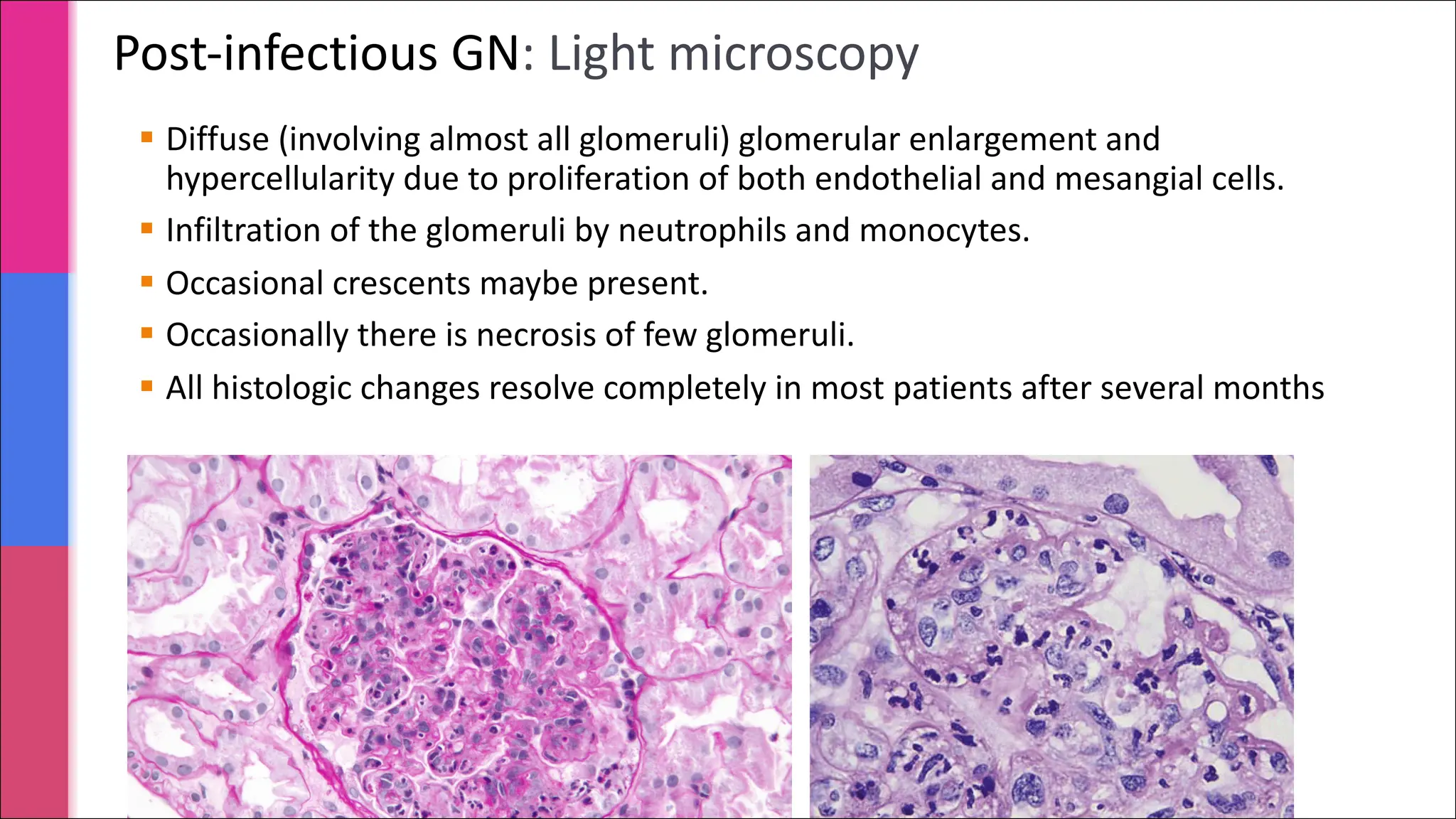
Post-infectious GN: Lightmicroscopy
§ Diffuse (involving almost all glomeruli) glomerular enlargement and
hypercellularity due to proliferation of both endothelial and mesangial cells.
§ Infiltration of the glomeruli by neutrophils and monocytes.
§ Occasional crescents maybe present.
§ Occasionally there is necrosis of few glomeruli.
§ All histologic changes resolve completely in most patients after several months
39.
https://www.stepwards.com/wp-
content/uploads/2016/01/j6npaxvA2gcDQTLLEm7mdQ_m.png
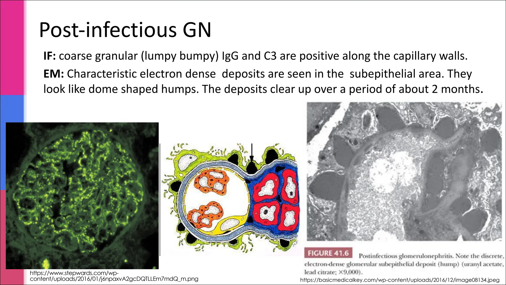
IF: coarse granular(lumpy bumpy) IgG and C3 are positive along the capillary walls.
EM: Characteristic electron dense deposits are seen in the subepithelial area. They
look like dome shaped humps. The deposits clear up over a period of about 2 months.
Post-infectious GN
https://basicmedicalkey.com/wp-content/uploads/2016/12/image08134.jpeg
40.
Post-infectious GN: clinicalfeatures
§ The onset of kidney disease is sudden.
§ The nephritic syndrome begins abruptly with oliguria, hematuria, facial
edema, hypertension and azotemia. The hematuria can be gross.
§ Serum C3 levels are low during the acute phase.
§ Diagnosis depends on serologic evidence of a rise in antibody titers to
streptococcal products e.g. ASO titre is positive.
§ This disease resolves in over 90% of patients. Rarely patients (usually adults)
develop progressive renal failure. Rarely children develop rapidly progressive
cresentric glomerulonephritis or chronic renal disease.
§ Grossly there is multiple punctate hemorrhagic spots on the kidney surface.
LUPUS NEPHROPATHY/ NEPHRITIS(LN)
§ Patients with the autoimmune disease called systemic lupus erythematosus (SLE)
tend to have renal involvement and it is known as lupus nephritis (LN).
§ It can present as nephrotic or nephritic syndrome.
§ LN is an immune complex mediated disease in which there is the deposition of
antigen antibody complexes in the glomeruli. The deposits trigger an inflammatory
response which in turn triggers the proliferation of the epithelial, endothelial and
mesangial cells of the glomeruli. It can even lead to glomerular necrosis.
§ The kidney in LN can show active lesions or chronic lesions or a combination of both.
§ Active lesions of the glomeruli include: endocapillary hypercellularity or extracapillary
proliferation (crescents), inflammation (glomerular or interstitial), fibrinoid necrosis
and subendothelial deposits.
§ Chronic lesions include: glomerular sclerosis, tubular atrophy and interstitial fibrosis.
§ The LN lesions have been classified into 6 classes by the International Society of
Nephrology/Renal Pathology Society (ISN/RPS). This classification helps give
information regarding the activity, chronicity and the prognosis of the disease.
43.
ADDITIONAL INFORMATION:
International Societyof Nephrology/Renal Pathology Society (ISN/RPS) classification of LN
The LN lesions have been classified into 6 classes. This classification helps give information
regarding the activity, chronicity and the prognosis of the disease. They are:
Class I/Minimal mesangial LN: no active or chronic lesions.
Class II/Mesangial Proliferative LN: no active or chronic lesions.
Class III/Focal LN: focal involvement of the glomeruli with active lesions or chronic lesions
or both a combination of both.
Class IV/Diffuse LN: is like class III but the involvement of the glomeruli is diffuse with
active lesions or chronic lesions or both a combination of both.
Class V/Membranous LN: same as membranous glomerulopathy. It may co-exist with Class
III or Class IV.
Class VI/Advanced sclerosing LN: is end stage kidney with no activity.
MPGN
§ It isa chronic progressive glomerulonephritis in older children and adults.
§ Histologically it is characterized by
1. mesangial hypercellularity with lobulation of glomerular tufts (lobular
accentuation of glomeruli)
2. and irregular thickening of the capillary wall due to the duplication or double
contouring of the GBM (also called as tram track lesions).
§ Patients may present with:
§ Nephrotic syndrome
§ Nephritic syndrome
§ Asymptomatic proteinuria
§ There are 2 main types: MPGN type I and type II (type II is also called as dense deposit
disease/DDD)
§ MPGN type I can be associated with Hepatitis B and C infection, SLE, infected
ventriculoatrial shunts and others.
§ IgA nephropathy(IgAN) is one of the most common type of primary
glomerulonephritis that presents as hematuria
§ IgAN is characterized by the deposition of IgA immunoglobulin in the
mesangium/ paramesangium of glomeruli.
§ It usually present as hematuria only and sometimes as nephritic
syndrome.
§ When it occurs in combination with vasculitis (leukocytoclastic vasculitis)
and multiorgan involvement then is referred to as Henoch-Schonlein
purpura.
Hematuria
48.
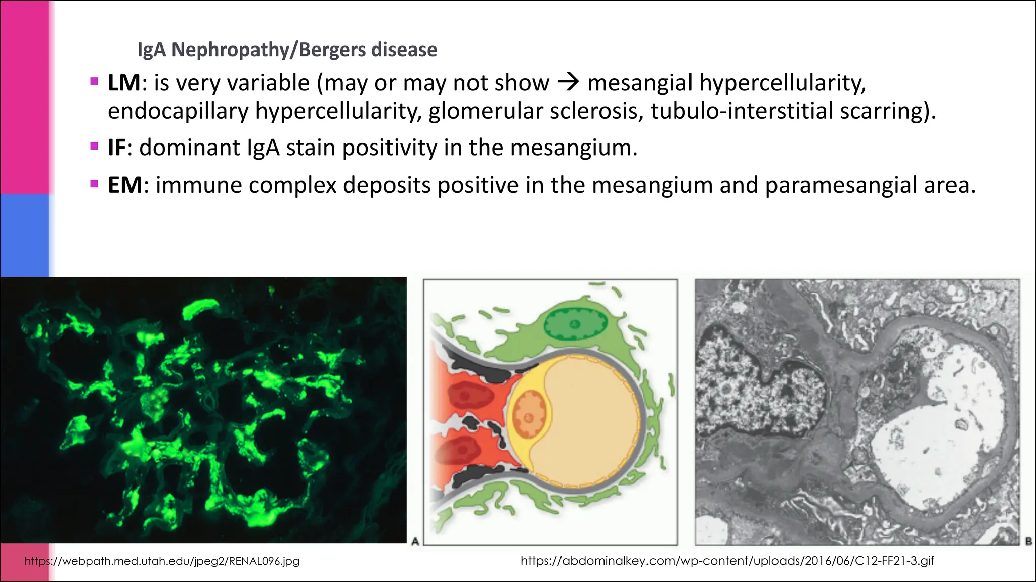
IgA Nephropathy/Bergers disease
§LM: is very variable (may or may not show à mesangial hypercellularity,
endocapillary hypercellularity, glomerular sclerosis, tubulo-interstitial scarring).
§ IF: dominant IgA stain positivity in the mesangium.
§ EM: immune complex deposits positive in the mesangium and paramesangial area.
https://webpath.med.utah.edu/jpeg2/RENAL096.jpg https://abdominalkey.com/wp-content/uploads/2016/06/C12-FF21-3.gif


![Nephrotic syndrome [full]](https://cdn.slidesharecdn.com/ss_thumbnails/nephroticsyndromefull-161026190255-thumbnail.jpg?width=640&height=640&fit=bounds)


